

Novel Radiographic Indexes for Elbow Stability Assessment: Part A-Cadaveric Validation
- PMID: 34306546
- PMCID: PMC8275710
- DOI: 10.1007/s43465-021-00407-4
Novel Radiographic Indexes for Elbow Stability Assessment: Part A-Cadaveric Validation
Abstract
Introduction: Elbow bony stability relies primarily on the high anatomic congruency between the humeral trochlea and the ulnar greater sigmoid notch. No practical tools are available to distinguish different morphotypes of the proximal ulna and herewith predict elbow stability. The aim of this study was to assess inter-observer reproducibility, evaluate diagnostic performance and determine responsiveness to change after simulated coronoid process fracture for three novel elbow radiographic indexes.
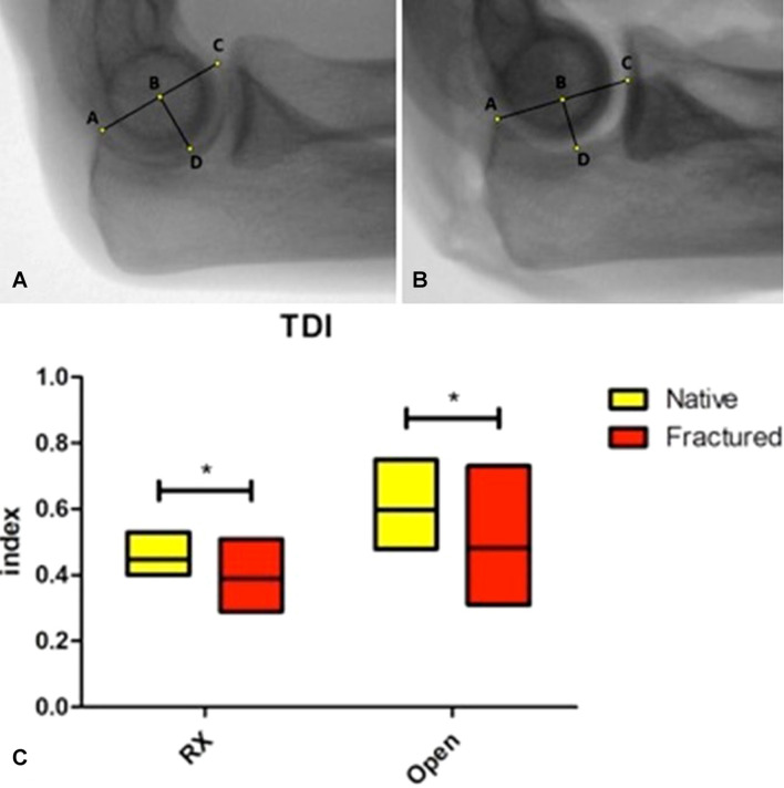
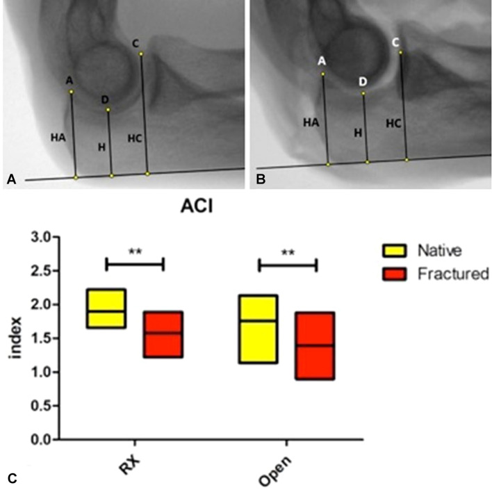
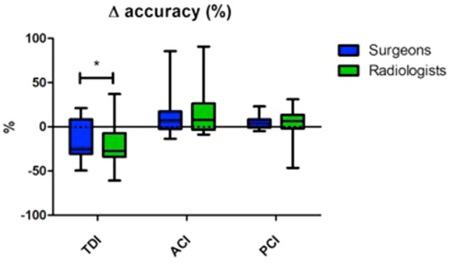
Methods: Ten fresh-frozen cadaver specimens of upper limbs from human donors were available for this study. Three primary indexes were defined, as well as two derived angles: Trochlear Depth Index (TDI); Posterior Coverage Index (PCI); Anterior Coverage Index (ACI); radiographic coverage angle (RCA); olecranon-diaphisary angle (ODA). Each index was first measured on standardized lateral radiographs and subsequently by direct measurement after open dissection. Finally, a type II coronoid fracture (Regan and Morrey classification) was created on each specimen and both radiographic and open measurements were repeated. All measurements were conducted by two orthopaedic surgeons and two dedicated musculoskeletal radiologists.
Results: All three indexes showed good or moderate inter-observer reliability and moderate accuracy and precision when compared to the gold standard (open measurement). A significant change between the radiographic TDI and ACI before and after simulated coronoid fracture was observed [TDI: decrease from 0.45 ± 0.03 to 0.39 ± 0.08 (p = 0.035); ACI: decrease from 1.90 ± 0.17 to 1.58 ± 0.21 (p = 0.001)]. As expected, no significant changes were documented for the PCI. Based on these data, a predictive model was generated, able to identify coronoid fractures with a sensitivity of 80% and a specificity of 100%.
Conclusion: New, simple and easily reproducible radiological indexes to describe the congruency of the greater sigmoid notch have been proposed. TDI and ACI change significantly after a simulated coronoid fracture, indicating a good responsiveness of these parameters to a pathological condition. Furthermore, combining TDI and ACI in a regression model equation allowed to identify simulated fractures with high sensitivity and specificity. The newly proposed indexes are, therefore, promising tools to improve diagnostic accuracy of coronoid fractures and show potential to enhance perioperative diagnostic also in cases of elbow instability and stiffness.
Level of evidence: Basic science study.
Clinical relevance: The newly proposed indexes are promising tools to improve diagnostic accuracy of coronoid fractures as well as to enhance perioperative diagnostic for elbow instability and stiffness.
Keywords: Cadaveric study; Elbow joint; Olecranon; Radiographic study; Ulna.
© The Author(s) 2021.
Conflict of interest statement
Conflict of interestAuthor FL declares that he has no conflict of interest. Author DC declares that he has no conflict of interest. Author ER declares that he has no conflict of interest. Author CEZ declares he has no conflict of interest. Author MV declares that he has no conflict of interest. Author LdG declares that he has no conflict of interest. Author AZ declares that he has no conflict of interest. Author MC declares that he has no conflict of interest. Author MBG declares that he has no conflict of interest. Author PA declares payment for development of educational presentations from Arthrex, outside the submitted work. Author PSR declares personal fees from Arthrex and Depuy (Johnson&Johnson), outside the submitted work.
Figures

References
-
- Tarassoli P, McCann P, Amirfeyz R. Complex instability of the elbow. Injury. 2017;48(3):568–577. - PubMed
-
- Lesman J, Luceri F, Domzalski M, Randelli PS, & Arrigoni P. The mini-open plication of medial collateral ligament of the elbow: a new arthroscopically assisted technique. https://journals.lww.com/shoulderelbowsurgery/Fulltext/2019/12000/The_Mi...
-
- Arrigoni P, Luceri F, Cucchi D, Tamini J, Randelli PS. Reliability of anterior medial collateral ligament plication of the elbow. Musculosk Surgery. 2018;1:19–102. - PubMed
-
- Rollo G, Luceri F, Pasquino A, Pichierri P, Tomarchio A, Bisaccia M, et al. Bone grafiting combined with Sauvé-Kapandji Procedures for the treatment of aseptic distal radius non-union. Journal of Biological Regulators and Homeostatic Agents. 2019;33(6 Suppl. 3):213–218. - PubMed
LinkOut - more resources
Full Text Sources
Research Materials
Miscellaneous