Case Reports
. 2021 Apr 30:11:e2021267.
doi: 10.4322/acr.2021.267. eCollection 2021.

Childhood pemphigus vulgaris is a challenging diagnosis

Affiliations
Case Reports

Childhood pemphigus vulgaris is a challenging diagnosis

Gastão Tenório Lins et al. Autops Case Rep. .

Abstract

Pemphigus Vulgaris (PV) is an uncommon autoimmune and blistering mucocutaneous disease. Childhood Pemphigus Vulgaris (CPV) is a pediatric variant of PV, which affects children below 12 years, being very rare among children under 10 years of age. CPV has similar clinical, histological, and immunological features as seen in PV in adults. The mucocutaneous clinical presentation is the most common in both age groups. Vesicles and erosions arising from the disease usually cause pain. A few CPV cases have been reported in the literature. This study reports a case of an 8-year-old male patient with oral lesions since the age of 3 years, and the diagnosis of pemphigus was achieved only 2 years after the appearance of the initial lesions. CPV remains a rare disease, making the diagnosis of this clinical case a challenge due to its age of onset and clinical features presented by the patient. Therefore, dentists and physicians should know how to differentiate CPV from other bullous autoimmune diseases more common in childhood.

Keywords: Child; Fluorescent Antibody Technique, Direct; Pemphigus.


Conflict of interest statement

Conflict of interest: The authors have no conflict of interest to declare.

Figures
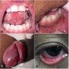
Figure 1
Figure 1. Lesions at the first appointment. A – Ulcerative lesions in the ventral surface of the tongue; B – Lesions with an ulcerative surface in lower labial mucosa (biopsy site); C – Ulcers in glans and foreskin; D – Ulcer in the mucosa of the lower eyelid.
Figure 2
Figure 2. Photomicrographs of the biopsy specimen. A – Intraepithelial blister with a single layer of basal cells recovering the conjunctive tissue and lymphoplasmacytic inflammatory infiltrate with eosinophils (H&E, 5X); B – High magnification of image A (H&E, 20X); C – Ulcered area with grouped acantholytic cells (*) with degenerative alterations (H&E, 40X); D – View of the inferior area of epithelial blister and suprabasal slit showing tiny area with rare basal cells still attached to the conjunctive tissue (H&E, 40X); E – Positive direct immunofluorescence showing IgG3 in the intercellular space of cells of the remaining epithelium basal layer on the conjunctiva.
Figure 3
Figure 3. Lesions on the last follow-up. A – Ulcerative lesions in the ventral surface of the tongue; B – Ulcers in the transition between alveolar mucosa and lower labial mucosa; C – Remission of lesions in glans and foreskin; D – Absence of lesion in the lower eyelid.

References

    1. Kincaid L, Weinstein M. Rituximab therapy for childhood pemphigus vulgaris. Pediatr Dermatol. 2016;33(2):e61–4. doi: 10.1111/pde.12744. - DOI - PubMed
    1. Surya V, Kumar P, Gupta S, Urs AB. Childhood pemphigus vulgaris: report of two cases with emphasis on diagnostic approach. Contemp Clin Dent. 2018;9(6) Suppl 2:S373–6. doi: 10.4103/ccd.ccd_461_18. - DOI - PMC - PubMed
    1. Gürcan HM, Mabrouk D, Razzaque Ahmed A. Management of pemphigus in pediatric patients. Minerva Pediatr. 2011;63(4):279–291. - PubMed
    1. Porro AM, Seque CA, Ferreira MCC, Enokihara MMSS. Pemphigus vulgaris. An Bras Dermatol. 2019;94(3):264–278. doi: 10.1590/abd1806-4841.20199011. - DOI - PMC - PubMed
    1. Gonul M, Keseroglu HO. Pediatric Phemphigus. Clin Pediatr Dermatol. 2015;1(1):1–3.

Publication types