

Cell Phone Selfies: Clinching the Diagnosis of Iris Microhemangiomatosis
- PMID: 34307330
- PMCID: PMC8280434
- DOI: 10.1159/000512343
Cell Phone Selfies: Clinching the Diagnosis of Iris Microhemangiomatosis
Abstract
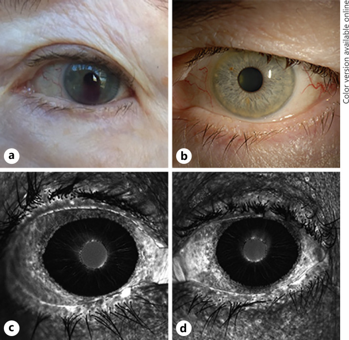
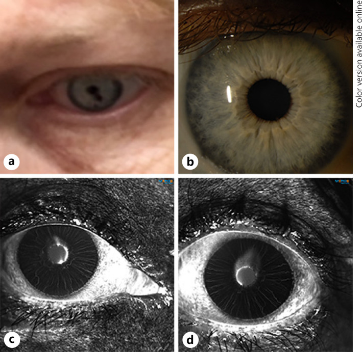
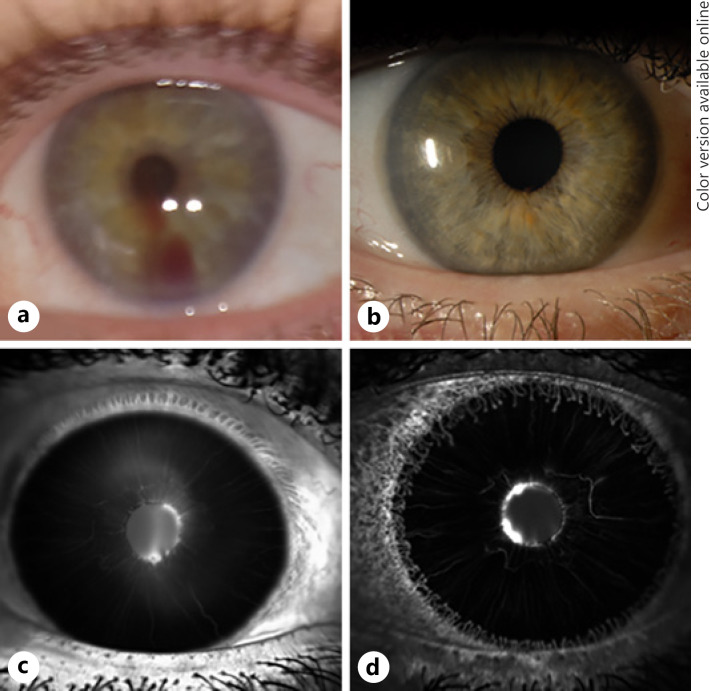
Four patients presented with a history of "blood" or a "dark spot" in the eye captured on cell phone photos. These episodes prompted presentation to an ophthalmologist where they all had normal slit lamp exams without a hyphema at initial evaluation. With evidence of a spontaneous hyphema seen on photos, further testing was performed including iris fluorescein angiography which revealed hyperfluorescent iris margin vascular tufts, confirming the diagnosis of iris microhemangiomatosis in each patient. All cases were managed conservatively, and only 1 needed topical antihypertensives. Without these patient-initiated photos, the diagnosis of iris microhemangiomatosis would likely remain elusive as slit lamp exam was normal at the time of initial examination in all 4 cases. Ophthalmologists should be aware of this rare diagnosis in the event a patient comes with a cell phone selfie documenting a spontaneous hyphema, especially when emanating from pupillary border.
Keywords: Hyphema; Microhemangiomatosis; Pathology of anterior segment; Selfie.
Copyright © 2021 by S. Karger AG, Basel.
Conflict of interest statement
The authors have no conflicts of interest to declare.
Figures

References
-
- Bennett TJ, Barry CJ. Ophthalmic imaging today: an ophthalmic photographer's viewpoint: a review. Clin Exp Ophthalmol. 2009;37((1)):2–13. - PubMed
-
- Hogarty DT, Hogarty JP, Hewitt AW. Smartphone use in ophthalmology: what is their place in clinical practice? Surv Ophthalmol. 2020;65((2)):250–62. - PubMed
Publication types
LinkOut - more resources
Full Text Sources
