. 2021 Jul 7:9:646113.
doi: 10.3389/fcell.2021.646113. eCollection 2021.

GLP-1-Induced AMPK Activation Inhibits PARP-1 and Promotes LXR-Mediated ABCA1 Expression to Protect Pancreatic β-Cells Against Cholesterol-Induced Toxicity Through Cholesterol Efflux

Affiliations

GLP-1-Induced AMPK Activation Inhibits PARP-1 and Promotes LXR-Mediated ABCA1 Expression to Protect Pancreatic β-Cells Against Cholesterol-Induced Toxicity Through Cholesterol Efflux

Rao Li et al. Front Cell Dev Biol. .

Abstract

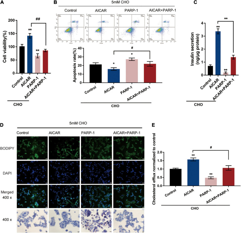
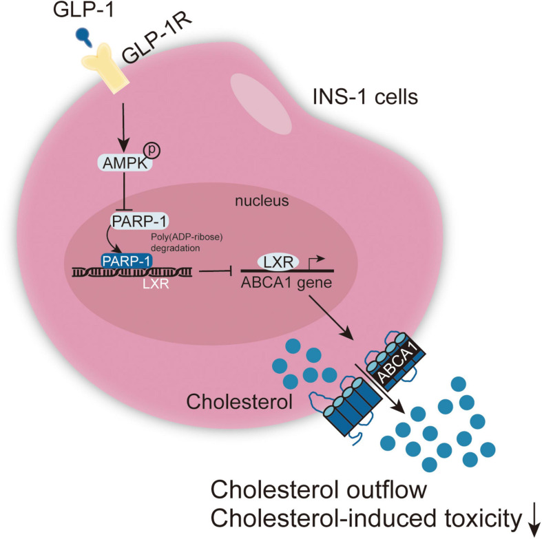
T2DM (Type 2 diabetes) is a complex, chronic disease characterized as insulin resistance and islet β-cell dysfunction. Bariatric surgeries such as Roux-en-Y gastric bypass (RYGB) surgery and laparoscopic sleeve gastrectomy (LSG) have become part of a critical treatment regimen in the treatment of obesity and T2DM. Moreover, GLP-1 increase following bariatric surgery has been regarded as a significant event in bariatric surgery-induced remission of T2DM. In this study, a high concentration cholesterol-induced lipotoxicity was observed in INS-1 cells, including inhibited cell viability and insulin secretion. Enhanced cell apoptosis and inhibited cholesterol efflux from INS-1 cells; meanwhile, ABCA1 protein level was decreased by cholesterol stimulation. Cholesterol-induced toxicity and ABCA1 downregulation were attenuated by GLP-1 agonist EX-4. GLP-1 induced AMPK phosphorylation during the protection against cholesterol-induced toxicity. Under cholesterol stimulation, GLP-1-induced AMPK activation inhibited PARP-1 activity, therefore attenuating cholesterol-induced toxicity in INS-1 cells. In INS-1 cells, PARP-1 directly interacted with LXR, leading to the poly(ADP-ribosyl)ation of LXRα and downregulation of LXR-mediated ABCA1 expression. In the STZ-induced T2DM model in rats, RYGB surgery or EX-4 treatment improved the glucose metabolism and lipid metabolism in rats through GLP-1 inhibition of PARP-1 activity. In conclusion, GLP-1 inhibits PARP-1 to protect islet β cell function against cholesterol-induced toxicity in vitro and in vivo through enhancing cholesterol efflux. GLP-1-induced AMPK and LXR-mediated ABCA1 expression are involved in GLP-1 protective effects.

Keywords: ABCA1; GLP-1; PARP-1; T2DM (type 2 diabetes); β-cell.


Conflict of interest statement

The authors declare that the research was conducted in the absence of any commercial or financial relationships that could be construed as a potential conflict of interest. The reviewer JY declared a shared affiliation with one of the authors LZ to the handling Editor at time of review.

Figures

FIGURE 1
FIGURE 1
GLP-1 downregulates ABCA1 expression and cholesterol accumulation-induced toxicity in INS-1 cells. INS-1 cells were cultured in a medium containing a series of concentrations of cholesterol (0, 1, 2.5, 5, or 10 mM) for 24 h and examined for (A) cell viability by MTT assay; (B) cell apoptosis by Flow cytometry; (C) the protein levels of ABCA1 by Immunoblotting. Next, INS-1 cells were cultured in a medium containing 0 or 5 mM cholesterol in the presence or absence of 10 nM Exendin-4 (EX-4) for 24 h and examined for (D) cell viability by MTT assay; (E) cell apoptosis by Flow cytometry; (F) insulin secretion by insulin ELISA kit; (G) the protein levels of ABCA1 by Immunoblotting; (H) intracellular cholesterol concentration using cholesterol assay kits; (I) intracellular lipid deposition by Oil Red O and BODIPY staining. (J) The cholesterol efflux from INS-1 cells by BODIPY-cholesterol assay; n = 3 for each experiment. *P < 0.05, **P < 0.01, ***P < 0.005, compared with control group, #P < 0.05, ##P < 0.01, compared with CHO + EX-4 group.
FIGURE 2
FIGURE 2
AMPK signaling is involved in GLP-1 regulation of ABCA1 expression and cholesterol-induced toxicity. (A) INS-1 cells were cultured in a medium containing 0 and 5 mM cholesterol in the presence or absence of 10 nM EX-4 for 24 h and examined for the protein levels of p-AMPK and AMPK by immunoblotting. Next, INS-1 cells were treated with EX-4 in the presence of AMPK activator AICAR or AMPK inhibitor Compound C under the stimulation of cholesterol and examined for (B) the protein levels of ABCA1 by immunoblotting; (C) cell viability by MTT assay; (D) cell apoptosis by Flow cytometry; (E) insulin secretion by insulin ELISA kit; (F) intracellular lipid deposition by Oil Red O and BODIPY staining. (G) The cholesterol efflux from INS-1 cells by BODIPY-cholesterol assay; n = 3 for each experiment. *P < 0.05, **P < 0.01, compared with the control group, #P < 0.05, ##P < 0.01 compared with CHO + EX-4 group, aP < 0.05, aaP < 0.01 compared with CHO + AICAR group, &&P < 0.01 compared with CHO + Compound C group.
FIGURE 3
FIGURE 3
GLP-1 regulates ABCA1 expression through AMPK-dependent inhibition on PARP1 activity. (A) Cholesterol-stimulated INS-1 cells were pre-treated with AMPK activator AICAR or AMPK inhibitor Compound C for 4 h before the addition of GLP-1 and incubated for another 24 h. The protein levels of PAR in INS-1 cell lysates were determined by immunoblotting. (B) PARP-1 knockdown was generated in INS-1 cells by the transfection of si-PARP-1. The transfection efficiency was confirmed by immunoblotting. (C) Cholesterol-stimulated INS-1 cells were transfected with si-PARP-1 or treated with PARP-1 inhibitor 3-AB and examined for the protein levels of ABCA1 by immunoblotting. (D) Cholesterol-stimulated INS-1 cells were treated with AMPK activator AICAR or PARP-1 alone or co-treated with AICAR and PARP-1 and examined for the protein levels of p-PARP-1 and ABCA1 by immunoblotting. **P < 0.01 compared with si-NC group or control group, ##P < 0.01 compared with AICAR group.
FIGURE 4
FIGURE 4
AMPK inhibits PARP1 activity to attenuate cholesterol-induced toxicity. Cholesterol-stimulated INS-1 cells were treated with AICAR or PARP-1 alone or co-treated with AICAR and PARP-1 and examined for (A) cell viability by MTT assay; (B) cell apoptosis by Flow cytometry; (C) insulin secretion by insulin ELISA kit; (D) intracellular lipid deposition by Oil Red O and BODIPY staining. (E) The cholesterol efflux from INS-1 cells by BODIPY-cholesterol assay. *P < 0.05, **P < 0.01 compared with control group, #P < 0.05, ##P < 0.01 compared with AICAR group.
FIGURE 5
FIGURE 5
PARP-1 inhibits LXR-induced ABCA1 transcription in INS-1 cells. (A) Confirmation of the interaction between LXR and PARP-1 in INS-1 cells using coimmunoprecipitation (Co-IP) assay. (B) LXRα poly(ADP-ribosyl)ation was detected in INS-1 by Poly(ADP-ribosyl)ation Assay. (C,D) INS-1 cells were transfected with si-PARP-1 in the presence or absence of LXR receptor agonist T0901317 and examined for the mRNA expression and protein levels of ABCA1 by real-time PCR and immunoblotting. n = 3 for each experiment. **P < 0.01 compared with the si-NC + DMSO group, #P < 0.05 compared with the si-NC + T0901317 group.
FIGURE 6
FIGURE 6
GLP-1 inhibits PARP-1 to improve islet function in T2DM rats. Sprague-Dawley (SD) rats were randomly assigned into five groups: non-treatment control group (n = 6), streptozotocin (STZ)-induced T2DM group (n = 6), T2DM plus sham surgery group (n = 7), T2DM plus RYGB surgery group (n = 9), and T2DM plus EX-4 treatment group (n = 8). At pre-operation (RYGB surgery or EX-4 injection), 1, 2, 4, and 8 weeks, the body weight (A), fasting blood glucose levels (FBG) in serum (B), the area under the curve (AUC) in serum for the OGTT (oral glucose tolerance test, mmol/L/hour) (C), total cholesterol (TC) in serum (D), triglyceride (TG) in serum (E), low-density lipoprotein cholesterol (LDL-C) in serum (F), and high-density lipoprotein cholesterol (HDL-C) in serum (G), GLP-1 levels in serum (H), and insulin levels in serum (I) were examined. (J,K) The TC and TG levels in pancreatic tissues of rats were determined. (L) The protein levels of p-AMPK, AMPK, PARP-1, LXRα, and ABCA1 were examined by immunoblotting in rats’ pancreatic tissues. n = 6, *P < 0.05, **P < 0.01 compared with NC group, #P < 0.05, ##P < 0.01 compared with T2DM + sham group, &P < 0.05, &&P < 0.01 compared with T2DM + RYGB group.
FIGURE 7
FIGURE 7
The pancreatic islet morphology and ABCA1 and GLP-1 protein levels. Sprague-Dawley (SD) rats were randomly assigned into five groups: non-treatment control group (n = 6), streptozotocin (STZ)-induced T2DM group (n = 6), T2DM plus sham surgery group (n = 7), T2DM plus RYGB surgery group (n = 9), and T2DM plus EX-4 treatment group (n = 8). (A) The histopathology of rats’ pancreatic tissues was examined by H&E staining. n = 6. (B,C) The protein contents and distribution of ABCA1 and PARP-1 were examined by IHC staining in rats’ pancreatic tissues. n = 6, *P < 0.05, **P < 0.01 compared with NC group, ##P < 0.01 compared with T2DM + sham group, &&P < 0.01 compared with T2DM + RYGB group.
FIGURE 8
FIGURE 8
A schematic graph of the mechanism. GLP-1 induces AMPK and LXR-mediated ABCA1 expression and inhibits PARP-1 to protect islet β cell function against cholesterol-induced toxicity in vitro and in vivo through enhancing cholesterol efflux.

References

    1. Allagnat F., Fukaya M., Nogueira T. C., Delaroche D., Welsh N., Marselli L., et al. (2012). /EBP homologous protein contributes to cytokine-induced pro-inflammatory responses and apoptosis in beta-cells. Cell Death Differ. 19 1836–1846. 10.1038/cdd.2012.67 - DOI - PMC - PubMed
    1. Andreone T. L., O’connor M., Denenberg A., Hake P., Zingarelli B. (2003). Poly(ADP-ribose) polymerase-1 regulates activation of activator protein-1 in murine fibroblasts. J. Immunol. 170 2113–2120. 10.4049/jimmunol.170.4.2113 - DOI - PubMed
    1. Bai P., Cantó C., Oudart H., Brunyánszki A., Cen Y., Thomas C., et al. (2011). PARP-1 inhibition increases mitochondrial metabolism through SIRT1 activation. Cell Metab. 13 461–468. 10.1016/j.cmet.2011.03.004 - DOI - PMC - PubMed
    1. Berchtold L. A., Prause M., Størling J., Mandrup-Poulsen T. (2016). Cytokines and Pancreatic beta-Cell Apoptosis. Adv. Clin. Chem. 75 99–158. 10.1016/bs.acc.2016.02.001 - DOI - PubMed
    1. Boni-Schnetzler M., Meier D. T. (2019). Islet inflammation in type 2 diabetes. Semin. Immunopathol. 41 501–513. 10.1007/s00281-019-00745-4 - DOI - PMC - PubMed