. 2021 Jul 8:9:693969.
doi: 10.3389/fcell.2021.693969. eCollection 2021.

Melatonin Protects Against Mdivi-1-Induced Abnormal Spindle Assembly and Mitochondrial Superoxide Production During Porcine Oocyte Maturation

Affiliations

Melatonin Protects Against Mdivi-1-Induced Abnormal Spindle Assembly and Mitochondrial Superoxide Production During Porcine Oocyte Maturation

Seul-Gi Yang et al. Front Cell Dev Biol. .

Abstract

Mitochondrial division inhibitor 1 (Mdivi-1) reportedly provides a close connection between oocyte maturation and mitochondrial function in pigs. N-acetyl-5-methoxy-tryptamine (melatonin) is known to be a representative antioxidant with the ability to rehabilitate meiotic maturation of porcine oocytes. However, the ability of melatonin to recover Mdivi-1-mediated disruption of spindle formation during meiotic maturation of porcine oocytes during in vitro maturation (IVM) has not been studied. Here, we first investigated changes in mitochondrial length, such as fragmentation and elongation form, in mature porcine oocytes during IVM. Mature oocytes require appropriate mitochondrial fission for porcine oocyte maturation. We identified a dose-dependent reduction in meiotic maturation in porcine oocytes following Mdivi-1 treatment (50, 75, and 100 μM). We also confirmed changes in mitochondrial fission protein levels [dynamin-related protein 1 phosphorylation at serine 616 (pDRP1-Ser616) and dynamin-related protein 1 (DRP1)], mitochondrial membrane potential, and ATP production in 75 μM Mdivi-1-treated oocytes. As expected, Mdivi-1 significantly reduced mitochondrial function and DRP1 protein levels and increased spindle abnormalities in porcine oocytes. In addition, we confirmed that melatonin restores abnormal spindle assembly and reduces meiotic maturation rates by Mdivi-1 during porcine oocyte maturation. Interestingly, the expression levels of genes that reduce DNA damage and improve tubulin formation were enhanced during porcine meiotic maturation. Taken together, these results suggest that melatonin has direct beneficial effects on meiotic maturation through tubulin formation factors during porcine oocyte maturation.

Keywords: Mdivi-1; melatonin; mitochondrial fission; pig oocyte maturation; spindle assembly.


Conflict of interest statement

The authors declare that the research was conducted in the absence of any commercial or financial relationships that could be construed as a potential conflict of interest.

Figures

FIGURE 1
FIGURE 1
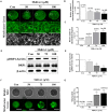
Changes of mitochondrial dynamics during meiotic maturation in porcine oocytes. (A–C) Porcine oocytes during meiotic maturation (0, 22, and 44 h) stained for mitochondrial fragmentation and elongation using MitoTracker green staining. Scale bar: 50 μm. (D,E) Representative Western blot and quantitative analyses of the expression of mitochondrial fission proteins [dynamin-related protein 1 phosphorylation at serine 616 (pDRP1-Ser616) and dynamin-related protein 1 (DRP1)] in immature and mature oocytes (0, 22, and 44 h). (F) Immunohistochemistry of DRP1 in various porcine ovarian follicles (1–2, 2–3, and 3–5 mm). Scale bar: 100 and 200 μm. Data in the bar graph represent the mean ± SD/SEM of three independent experiments (p < 0.05; ∗∗p < 0.01).
FIGURE 2
FIGURE 2
Effects of mitochondrial division inhibitor 1 (Mdivi-1) exposure on mitochondrial dynamics and distribution during meiotic maturation in porcine oocytes. (A–C) Representative images of mitochondrial length (fragmentation and elongation) in 0, 50, 75, or 100 μM Mdivi-1-treated oocytes. Scale bar: 50 μm. (D,E) Representative Western blot of the expression of mitochondrial fission proteins [dynamin-related protein 1 phosphorylation at serine 616 (pDRP1-Ser616) and dynamin-related protein 1 (DRP1)] in control and Mdivi-1-treated oocytes. Here, β-actin was used as a loading control. The ratios of phosphorylated/total proteins were calculated and plotted in all experimental groups. (F,G) Ratios of mitochondria aggregation were detected using MitoTracker green. Scale bar: 200 μm. Data represent the mean ± SD/SEM of three biological replicates (p < 0.05; ∗∗p < 0.01; ∗∗∗p < 0.001).
FIGURE 3
FIGURE 3
Effects of mitochondrial division inhibitor 1 (Mdivi-1) exposure on mitochondrial functions in mature porcine oocytes. (A–C) Representative images of intracellular and mitochondrial reactive oxygen species (ROS) levels in control, 50, 75, or 100 μM Mdivi-1-treated oocytes. Green: intracellular ROS; red: mitochondria-derived superoxide. Scale bar: 200 μm. (D,E) Typical images of mitochondrial membrane potential (MMP) in the control and Mdivi-1 (50, 75, and 100 μM) groups. Scale bar: 100 μm. (F) Relative ATP contents of mature porcine oocytes from various Mdivi-1-treated groups. Data in the bar graph represent the mean ± SD of three independent experiments (p < 0.05; ∗∗p < 0.01; ∗∗∗p < 0.001).
FIGURE 4
FIGURE 4
Effects of mitochondrial division inhibitor 1 (Mdivi-1) on spindle assembly in mature porcine oocytes. (A,B) Representative images of mitochondrial activation in the control and Mdivi-1-treated groups. Orange: mitochondria; blue: chromosome. Scale bar: 50 μm. (B) Quantitative analysis of mitochondria activation in the control and Mdivi-1-treated groups. (C) Representative images of spindle morphology and chromosome alignment in the control and Mdivi-1-treated groups. Green: α-tubulin; blue: chromosome. Scale bar: 10 μm. Abnormal spindle (D) and misaligned chromosome (E) rates of mature oocytes in the control and Mdivi-1-treated groups. (F) Arrangement of spindle assembly in the control and Mdivi-1-treated groups. Green: α-tubulin; blue: chromosome. Data represent the mean ± SD of three biological replicates (∗∗p < 0.01; ∗∗∗p < 0.001).
FIGURE 5
FIGURE 5
Effects of mitochondrial division inhibitor 1 (Mdivi-1) on the DNA damage and tubulin formation-related genes in mature porcine oocytes. (A) Detection of RAD51 (green fluorescence), γ-H2AX (red fluorescence), and chromosome (blue fluorescence) expression using immunofluorescence (IF) staining in mature porcine oocytes. Scale bar: 50 μm. (B) Percentage of γ-H2AX-positive cell ratios in mature porcine oocytes. (C,D) Expression levels of DNA damage-related genes (Rad51 and γ-H2ax) after Mdivi-1 treatment in mature porcine oocytes. (E) The expression level of melatonin synthesis enzyme gene (Aanat) after Mdivi-1 treatment in mature porcine oocytes. (F–H) Expression levels of tubulin formation-related genes (Tuba1a, Atat1, and Map2) after Mdivi-1 treatment in mature porcine oocytes. Data in the bar graph represent the mean ± SEM of three independent experiments (p < 0.05; ∗∗p < 0.01).
FIGURE 6
FIGURE 6
Effect of melatonin on mitochondrial fission and spindle assembly in mature porcine oocytes. (A,B) Representative Western blot of the mitochondrial fission [dynamin-related protein 1 phosphorylation at serine 616 (pDRP1-Ser616) and dynamin-related protein 1 (DRP1)] protein levels in the control, mitochondrial division inhibitor 1 (Mdivi-1), melatonin, and Mdivi-1+melatonin groups. Here, β-actin was used as a loading control. The ratios of phosphorylated/total proteins were calculated and plotted in all experimental groups. (C) Representative images of spindle morphology and chromosome alignment in the control, Mdivi-1, melatonin, and Mdivi-1 + melatonin groups. Green: α-tubulin; blue: chromosome. Scale bar: 10 μm. Abnormal spindle (D) and misaligned chromosome (E) rates of mature oocytes in the control, Mdivi-1, melatonin, and Mdivi-1 + melatonin groups. (F) Arrangement of spindle assembly in the control, Mdivi-1, melatonin, and Mdivi-1 + melatonin groups. Green: α-tubulin; blue: chromosome. Data represent the mean ± SD/SEM of three biological replicates (p < 0.05; ∗∗p < 0.01; ∗∗∗p < 0.001).
FIGURE 7
FIGURE 7
Effects of melatonin on the intracellular and mitochondrial reactive oxygen species (ROS) and tubulin formation-related protein in mature porcine oocytes. (A–C) Fluorescence intensity of intracellular (green) and mitochondrial (red) ROS using DCF-DA and Mito-SOX staining. Scale bar: 100 μm. (D,E) Representative Western blot of the AC-TUBULIN protein level in the control, mitochondrial division inhibitor 1 (Mdivi-1), melatonin, and Mdivi-1 + melatonin groups. Here, β-actin was used as a loading control. Data in the bar graph represent the mean ± SEM of three independent experiments (p < 0.05; ∗∗∗p < 0.001).
FIGURE 8
FIGURE 8
Effects of melatonin on the DNA damage and tubulin formation-related genes in mature porcine oocytes. (A) Immunofluorescence of RAD51 and γ-H2AX protein expression of mature porcine oocytes. Scale bar: 100 μm. (B) Percentage of γ-H2AX-positive cell ratios in the control, mitochondrial division inhibitor 1 (Mdivi-1), melatonin, and Mdivi-1 + melatonin groups of mature porcine oocytes. (C,D) Expression levels of DNA damage-related genes (Rad51 and γ-H2ax) in the control, Mdivi-1, melatonin, and Mdivi-1 + melatonin groups. (E) Expression levels of the melatonin synthesis enzyme gene (Aanat) in the control, Mdivi-1, melatonin, and Mdivi-1 + melatonin groups. (F–H) Expression levels of tubulin formation-related genes (Tuba1a, Atat1, and Map2) in the control, Mdivi-1, melatonin, and Mdivi-1 + melatonin groups. Data in the bar graph represent the mean ± SEM of three independent experiments (p < 0.05).
FIGURE 9
FIGURE 9
Schematic diagram indicating the suggested protective effects of melatonin on Mdivi-1-inhibited mitochondrial fission in porcine oocytes. In non-treated conditions, mitochondrial fission and fragmentation (<1 μm) increased during oocyte maturation. The Mdivi-1-exposed oocyte increased abnormal spindle assembly (Mdivi-1-induced damage, 1) and mitochondria-derived superoxide assembly (Mdivi-1-induced damage, 2) by blocked mitochondrial fission in pig. Melatonin enhances oocyte maturation capacity against Mdivi-1-mediated damages through restoration of spindle assembly (melatonin recovery effect, 1) and anti-oxidative effector (melatonin recovery effect, 1). Melatonin improves meiotic maturation in Mdivi-1-exposed oocytes as a recovery and protective effect of mitochondrial superoxide and abnormal spindle assembly during IVM.

Similar articles

Cited by

References

    1. Adam-Zahir S., Plowman P. N., Bourton E. C., Sharif F., Parris C. N. (2014). Increased γ-H2AX and Rad51 DNA repair biomarker expression in human cell lines resistant to the chemotherapeutic agents nitrogen mustard and cisplatin. Chemotherapy 60 310–320. 10.1159/000430086 - DOI - PubMed
    1. Adhikari D., Liu K. (2014). The regulation of maturation promoting factor during prophase I arrest and meiotic entry in mammalian oocytes. Mol. Cell. Endocrinol. 382 480–487. 10.1016/j.mce.2013.07.027 - DOI - PubMed
    1. An Q., Peng W., Cheng Y., Lu Z., Zhou C., Zhang Y., et al. (2019). Melatonin supplementation during in vitro maturation of oocyte enhances subsequent development of bovine cloned embryos. J. Cell. Physiol. 234 17370–17381. 10.1002/jcp.28357 - DOI - PubMed
    1. Bennabi I., Terret M.-E., Verlhac M.-H. (2016). Meiotic spindle assembly and chromosome segregation in oocytes. J. Cell Biol. 215 611–619. 10.1083/jcb.201607062 - DOI - PMC - PubMed
    1. Bordt E. A., Clerc P., Roelofs B. A., Saladino A. J., Tretter L., Adam-Vizi V., et al. (2017). The putative Drp1 inhibitor mdivi-1 is a reversible mitochondrial complex I inhibitor that modulates reactive oxygen species. Developmental cell 40 583–594.e6. - PMC - PubMed