

Two and Three-Dimensional Echocardiography in Primary Mitral Regurgitation: Practical Hints to Optimize the Surgical Planning
- PMID: 34307510
- PMCID: PMC8295501
- DOI: 10.3389/fcvm.2021.706165
Two and Three-Dimensional Echocardiography in Primary Mitral Regurgitation: Practical Hints to Optimize the Surgical Planning
Abstract
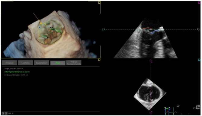
Primary mitral regurgitation (MR) is the second most common valvular disease, characterized by a high burden in terms of quality of life, morbidity, and mortality. Surgical treatment is considered the best therapeutic strategy for patients with severe MR, especially if they are symptomatic. However, pre-operative echocardiographic evaluation is an essential step not only for surgical candidate selection but also to avoid post-operative complications. Therefore, a strong collaboration between cardiologists and cardiac surgeons is fundamental in this setting. A meticulous pre-operative echocardiographic exam, both with transthoracic or transesophageal echocardiography, followed by a precise report containing anatomical information and parameters should always be performed to optimize surgical planning. Moreover, intraoperative transesophageal evaluation is often required by cardiac surgeons as it may offer additive important information with different hemodynamic conditions. Three-dimensional echocardiography has recently gained higher consideration and availability for the evaluation of MR, providing more insights into mitral valve geometry and MR mechanism. This review paper aims to realize a practical overview on the main use of basic and advanced echocardiography in MR surgical planning and to provide a precise checklist with reference parameters to follow when performing pre-operative echocardiographic exam, in order to aid cardiologists to provide a complete echocardiographic evaluation for MR operation planning from clinical and surgical point-of-view.
Keywords: echocardiography; mitral regurgitation; planning; surgery; three-dimensional.
Copyright © 2021 Pastore, Mandoli, Sannino, Dokollari, Bisleri, D'Ascenzi, Cavigli, Pasquini, Lisi, Ghionzoli, Santoro, Miglioranza, Focardi, Patti, Valente, Mondillo and Cameli.
Conflict of interest statement
The authors declare that the research was conducted in the absence of any commercial or financial relationships that could be construed as a potential conflict of interest.
Figures


References
-
- Nishimura RA, Otto CM, Bonow RO, Carabello BA, Erwin JP, III, Fleisher LA, et al. . 2017 AHA/ACC focused update of the 2014 AHA/ACC guideline for the management of patients with valvular heart disease: a report of the American College of Cardiology/American Heart Association Task Force on Clinical Practice Guidelines. Circulation. (2017) 135:e1159–95. 10.1161/CIR.0000000000000503 - DOI - PubMed
-
- Lang RM, Badano LP, Mor-Avi V, Afilalo J, Armstrong A, Ernande L, et al. . Recommendations for cardiac chamber quantification by echocardiography in adults: an update from the American Society of Echocardiography and the European Association of Cardiovascular Imaging. Eur Heart J Cardiovasc Imaging. (2015) 16:233–70. 10.1093/ehjci/jev014 - DOI - PubMed
-
- Yazdchi F, Kaneko T, Tang GHL. Surgical versus percutaneous approaches for degenerative mitral valve repair: a review. Struct. Heart. (2019) 3:176–84. 10.1080/24748706.2019.1596337 - DOI
Publication types
LinkOut - more resources
Full Text Sources