

Mycobacterial Infection of Precision-Cut Lung Slices Reveals Type 1 Interferon Pathway Is Locally Induced by Mycobacterium bovis but Not M. tuberculosis in a Cattle Breed
- PMID: 34307535
- PMCID: PMC8299756
- DOI: 10.3389/fvets.2021.696525
Mycobacterial Infection of Precision-Cut Lung Slices Reveals Type 1 Interferon Pathway Is Locally Induced by Mycobacterium bovis but Not M. tuberculosis in a Cattle Breed
Abstract
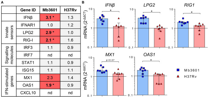
Tuberculosis exacts a terrible toll on human and animal health. While Mycobacterium tuberculosis (Mtb) is restricted to humans, Mycobacterium bovis (Mb) is present in a large range of mammalian hosts. In cattle, bovine TB (bTB) is a noticeable disease responsible for important economic losses in developed countries and underestimated zoonosis in the developing world. Early interactions that take place between mycobacteria and the lung tissue early after aerosol infection govern the outcome of the disease. In cattle, these early steps remain poorly characterized. The precision-cut lung slice (PCLS) model preserves the structure and cell diversity of the lung. We developed this model in cattle in order to study the early lung response to mycobacterial infection. In situ imaging of PCLS infected with fluorescent Mb revealed bacilli in the alveolar compartment, in adjacent or inside alveolar macrophages, and in close contact with pneumocytes. We analyzed the global transcriptional lung inflammation signature following infection of PCLS with Mb and Mtb in two French beef breeds: Blonde d'Aquitaine and Charolaise. Whereas, lungs from the Blonde d'Aquitaine produced high levels of mediators of neutrophil and monocyte recruitment in response to infection, such signatures were not observed in the Charolaise in our study. In the Blonde d'Aquitaine lung, whereas the inflammatory response was highly induced by two Mb strains, AF2122 isolated from cattle in the UK and Mb3601 circulating in France, the response against two Mtb strains, H37Rv, the reference laboratory strain, and BTB1558, isolated from zebu in Ethiopia, was very low. Strikingly, the type I interferon pathway was only induced by Mb but not Mtb strains, indicating that this pathway may be involved in mycobacterial virulence and host tropism. Hence, the PCLS model in cattle is a valuable tool to deepen our understanding of early interactions between lung host cells and mycobacteria. It revealed striking differences between cattle breeds and mycobacterial strains. This model could help in deciphering biomarkers of resistance vs. susceptibility to bTB in cattle as such information is still critically needed for bovine genetic selection programs and would greatly help the global effort to eradicate bTB.
Keywords: Mycobacterium bovis; alveolar macrophages; cattle; ex vivo; precision cut lung slices; type I interferon.
Copyright © 2021 Remot, Carreras, Coupé, Doz-Deblauwe, Boschiroli, Browne, Marquant, Descamps, Archer, Aseffa, Germon, Gordon and Winter.
Conflict of interest statement
The authors declare that the research was conducted in the absence of any commercial or financial relationships that could be construed as a potential conflict of interest. The handling editor declared a past collaboration with one of the authors SG.
Figures


Similar articles
-
Innate cytokine profiling of bovine alveolar macrophages reveals commonalities and divergence in the response to Mycobacterium bovis and Mycobacterium tuberculosis infection.Tuberculosis (Edinb). 2014 Jul;94(4):441-50. doi: 10.1016/j.tube.2014.04.004. Epub 2014 May 5. Tuberculosis (Edinb). 2014. PMID: 24882682
-
Comparative 'omics analyses differentiate Mycobacterium tuberculosis and Mycobacterium bovis and reveal distinct macrophage responses to infection with the human and bovine tubercle bacilli.Microb Genom. 2018 Mar;4(3):e000163. doi: 10.1099/mgen.0.000163. Epub 2018 Mar 20. Microb Genom. 2018. PMID: 29557774 Free PMC article.
-
Genetic characterization of the Blonde d'Aquitaine cattle breed using microsatellite markers and relationship with three other French cattle populations.J Anim Breed Genet. 2011 Jun;128(3):201-8. doi: 10.1111/j.1439-0388.2010.00890.x. Epub 2011 Jan 12. J Anim Breed Genet. 2011. PMID: 21554414
-
Epidemiology of Mycobacterium bovis and Mycobacterium tuberculosis in animals: Transmission dynamics and control challenges of zoonotic TB in Ethiopia.Prev Vet Med. 2018 Oct 1;158:1-17. doi: 10.1016/j.prevetmed.2018.06.012. Epub 2018 Jun 28. Prev Vet Med. 2018. PMID: 30220382 Review.
-
[Development of antituberculous drugs: current status and future prospects].Kekkaku. 2006 Dec;81(12):753-74. Kekkaku. 2006. PMID: 17240921 Review. Japanese.
Cited by
-
The Activation of the RIG-I/MDA5 Signaling Pathway upon Influenza D Virus Infection Impairs the Pulmonary Proinflammatory Response Triggered by Mycoplasma bovis Superinfection.J Virol. 2023 Feb 28;97(2):e0142322. doi: 10.1128/jvi.01423-22. Epub 2023 Jan 24. J Virol. 2023. PMID: 36692289 Free PMC article.
-
Punch-excised explants of bovine mammary gland to model early immune response to infection.J Anim Sci Biotechnol. 2023 Jul 7;14(1):100. doi: 10.1186/s40104-023-00899-0. J Anim Sci Biotechnol. 2023. PMID: 37420291 Free PMC article.
-
Respiratory bioenergetics is enhanced in human, but not bovine macrophages after exposure to M. bovis PPD: Exploratory insights into overall similar Cellular Metabolic Profiles.Innate Immun. 2024 Aug;30(6-8):136-149. doi: 10.1177/17534259241296630. Epub 2024 Nov 20. Innate Immun. 2024. PMID: 39563509 Free PMC article.
-
Hyper-induction of IL-6 after TLR1/2 stimulation in calves with bovine respiratory disease.PLoS One. 2024 Nov 14;19(11):e0309964. doi: 10.1371/journal.pone.0309964. eCollection 2024. PLoS One. 2024. PMID: 39541407 Free PMC article.
-
A bovine pulmosphere model and multiomics reveal early host response signature in tuberculosis.Commun Biol. 2025 Apr 4;8(1):559. doi: 10.1038/s42003-025-07883-6. Commun Biol. 2025. PMID: 40186000 Free PMC article.
References
LinkOut - more resources
Full Text Sources
Miscellaneous