

Collagen VI-related myopathy with scoliosis alone: A case report and literature review
- PMID: 34307582
- PMCID: PMC8283577
- DOI: 10.12998/wjcc.v9.i19.5302
Collagen VI-related myopathy with scoliosis alone: A case report and literature review
Abstract
Background: Scoliosis is a complex three-dimensional deformity of spine and one of the common complications of collagen VI-related myopathy, caused by mutations in collagen type VI alpha 1 chain (COL6A1), COL6A2, and COL6A3 genes. The typical clinical presentations of collagen VI-related myopathy include weakness, hypotonia, laxity of distal joints, contractures of proximal joints, and skeletal deformities.
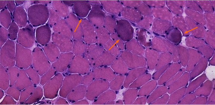
Case summary: A 28-year-old female presented with scoliosis for 28 years without weakness, hypotonia, laxity of distal joints, and contracture of proximal joints. Computed tomography and magnetic resonance imaging revealed hemivertebra, butterfly vertebra, and the missing vertebral space. Patients underwent orthopedic surgery and paravertebral muscle biopsy. The Cobb angle dropped from 103.4° to 52.9°. However, the muscle biopsy showed neurogenic muscular atrophy with myogenic lesions, suggesting congenital muscular dystrophy. Gene analysis indicated that mutations in COL6A1 (c.1612-10G>A) and COL6A2 (c.115+10G>T, c.2749G>A). Immunohistochemistry staining for collagen VI displayed shallow and discontinuous. Eventually, the patient was diagnosed as collagen VI-related myopathy.
Conclusion: This newly found subtype of collagen VI-related myopathy has no typical manifestations; however, it is characterized by severe scoliosis and congenital vertebral deformity.
Keywords: Case report; Collagen VI-related myopathy; Genetic testing; Neuromuscular diseases; Paravertebral pathology; Scoliosis.
©The Author(s) 2021. Published by Baishideng Publishing Group Inc. All rights reserved.
Conflict of interest statement
Conflict-of-interest statement: The authors declare that they have no conflict of interest.
Figures


Similar articles
-
[Clinical manifestations and genetics analysis of collagen type Ⅵ-related myopathy caused by variants in COL6A3 gene].Zhonghua Er Ke Za Zhi. 2019 Feb 2;57(2):136-141. doi: 10.3760/cma.j.issn.0578-1310.2019.02.014. Zhonghua Er Ke Za Zhi. 2019. PMID: 30695889 Chinese.
-
[Collagen VI-related muscle disorders].Brain Nerve. 2011 Nov;63(11):1169-78. Brain Nerve. 2011. PMID: 22068469 Review. Japanese.
-
Bethlem Myopathy (Collagen VI-Related Dystrophies): A Retrospective Cohort Study on Musculoskeletal Pathologies and Clinical Course.J Pediatr Orthop. 2023 Feb 1;43(2):e163-e167. doi: 10.1097/BPO.0000000000002283. Epub 2022 Oct 26. J Pediatr Orthop. 2023. PMID: 36607927
-
COL6A1 mutation leading to Bethlem myopathy with recurrent hematuria: a case report.BMC Neurol. 2019 Feb 26;19(1):32. doi: 10.1186/s12883-019-1263-0. BMC Neurol. 2019. PMID: 30808312 Free PMC article.
-
A novel de novo COL6A1 mutation emphasizes the role of intron 14 donor splice site defects as a cause of moderate-progressive form of ColVI myopathy - a case report and review of the genotype-phenotype correlation.Folia Neuropathol. 2017;55(3):214-220. doi: 10.5114/fn.2017.70486. Folia Neuropathol. 2017. PMID: 28984114 Review.
Cited by
-
Suspected malnutrition-induced reversible feline skin fragility syndrome in a cat with congenital axial deformities.Can Vet J. 2024 Mar;65(3):227-233. Can Vet J. 2024. PMID: 38434166 Free PMC article.
References
-
- Shakil H, Iqbal ZA, Al-Ghadir AH. Scoliosis: review of types of curves, etiological theories and conservative treatment. J Back Musculoskelet Rehabil. 2014;27:111–115. - PubMed
-
- Pahys JM, Guille JT. What's New in Congenital Scoliosis? J Pediatr Orthop. 2018;38:e172–e179. - PubMed
-
- McMaster MJ, Ohtsuka K. The natural history of congenital scoliosis. A study of two hundred and fifty-one patients. J Bone Joint Surg Am. 1982;64:1128–1147. - PubMed
Publication types
LinkOut - more resources
Full Text Sources
Miscellaneous
