Review
. 2021 Jul 14:8:100369.
doi: 10.1016/j.ejro.2021.100369. eCollection 2021.

Spinal vascular lesions: anatomy, imaging techniques and treatment

Affiliations
Review

Spinal vascular lesions: anatomy, imaging techniques and treatment

Valerio Da Ros et al. Eur J Radiol Open. .

Abstract

Background: Vascular lesions of the spinal cord are rare but potentially devastating conditions whose accurate recognition critically determines the clinical outcome. Several conditions lead to myelopathy due to either arterial ischemia, venous congestion or bleeding within the cord. The clinical presentation varies, according with the different aetiology and mechanism of damage.

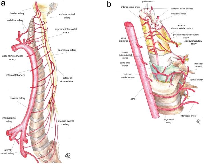
Purpose: The aim is to provide a comprehensive review on the radiological features of the most common vascular myelopathies, passing through the knowledge of the vascular spinal anatomy and the clinical aspects of the different aetiologies, which is crucial to promptly address the diagnosis and the radiological assessment.

Keywords: MRI; Neuroimaging; Spinal angiography; Spinal cord anatomy; Vascular myelopathies; Vascular spinal cord lesions.


Conflict of interest statement

The authors declare that they have no conflict of interest.

Figures

Fig. 1
Fig. 1
Cross sectional MRI anatomy of the spinal cord in cervical segment. The grey matter appears hyperintense on T2 sequences and its H-shaped borders the three white matter columns: the anterior column (yellow dotted line), the lateral column (green dotted line) and the dorsal column (red dotted line) (For interpretation of the references to colour in this figure legend, the reader is referred to the web version of this article).
Fig. 2
Fig. 2
(a and b). Sectional representation of the arterial blood supply and feeling vessels to spinal cord (see the text for further details), whose catheterization is mandatory during spinal angiography. Along the cranio-caudal direction, the angiographic study should include and demonstrate: vertebral arteries, ascending cervical arteries, deep cervical arteries (not shown), supreme intercostal arteries, intercostal arteries, lumbar arteries, median and lateral sacral arteries. Identification of the artery of Adamkiewicz is essential, because spinal vascular malformations might themselves originate from this major feeder of the anterior spinal artery.
Fig. 3
Fig. 3
Sectional representation of the spinal cord venous system (see the text for further details).
Fig. 4
Fig. 4
T2 weighted images on axial plane in the same patient; a) T2-TSE sequence and b) T2-GRE sequence. To note the better identification of central grey matter in cervical spine in GRE than TSE sequence related to lower sensitivity of the pulsation artifact for the GRE sequence in the cervical spinal cord.
Fig. 5
Fig. 5
Sagittal T2-TSE (a) and Diffusion Weighted (b) sequences in a 74 years old female patient with acute spinal ischemia. Axial T2-TSE sequence on axial plane (c) at level D12-L1. Diffusion weighted sequence clearly shows the restricted diffusion of the dorsal spinal cord (white brackets in b) corresponding to the hyperintense signal in the sagittal T2 sequence (white brackets in a); T2 sequences depicted a moderately swollen appearance of the medullary conus and the full involvement (grey and white matter) of the spinal cord.
Fig. 6
Fig. 6
Spinal dural arteriovenous fistula (SDAVF): a low-flow shunt between a radiculomeningeal artery and a medullary vein inside the dura mater results in vein arterialization and retrograde blood flow into a congested coronal venous plexus.
Fig. 7
Fig. 7
Contrast enhanced CT in in a 65-year-old man with accidental fall and progressive lower limb weakness. Sagittal (a, b) and coronal (c) images demonstrate the early appearance of a serpiginous vascular structure along the posterior profile of the spinal cord between D10 and D11. Axial scan (d) confirms the posterior peri-medullary anomalous vascular structure characterized by early enhancement after administration of contrast medium (white arrows in a, b, c and d).
Fig. 8
Fig. 8
MRI of the same patient in Fig. 7. Sagittal T2-TSE weighted (a) and T1-TSE weighted (b) images; axial T2-TSE weighted images at D7 (c) and D11 (d). T2 sequences show abnormal hyperintensity and swollen appearance of the dorsal spinal cord with cross-sectional involvement (bracket in a, white arrows in c and d). Vertebral haemangioma in D5 (white dotted line in a).
Fig. 9
Fig. 9
Spinal dural arteriovenous fistula (SDAVF). Microcatheterism (A) and 3D reconstruction (B) following injection of the right L3 segmental artery show an anomalous arteriovenous shunt between a radiculomeningeal artery and a medullary vein, located underneath the vertebral pedicle.
Fig. 10
Fig. 10
Spinal epidural arterovenous fistula (SEDAVF) with peri-medullary drainage: the arteriovenous shunt is located in the epidural space and consists of a large dilated extradural venous pouch; spinal and paraspinal veins arise from the pouch, leading to spinal cord congestion.
Fig. 11
Fig. 11
MRI in a 58-year-old man suffering from myelopathic symptoms in the past 18 months. Sagittal T2-weighted (a), T1-weighted (b) and contrast-enhanced T1-weighted (c) MRI images demonstrate enlargement and inhomogeneous T2 hyperintensity of the spinal cord between D8 and L1 (white bracket in a) with vanished enhancement after administration of gadolinium-based contrast medium (white bracket in c). Axial T2-weighted image (d) at level of D10 confirms the cross-sectional involvement of the spinal cord (white arrow).
Fig. 12
Fig. 12
Spinal epidural arteriovenous fistula (SEDAVF) with intradural venous drainage of the same patient in Fig. 11. Digital subtraction angiography (A) and 3D reconstruction (B) following injection of the right L2 segmental artery demonstrate a venous pouch in the epidural space and an intradural draining vein.
Fig. 13
Fig. 13
Follow-up MRI scan of the same patient in Fig. 11, Fig. 12. Sagittal (a) and axial (b, c) T2-weighted images show the severe reduction of dorsal spinal cord thickness (white bracket in a). Axial images demonstrate mild hyperintensity in the posterior median column at D8 (black arrow in b); to note the severe thinning and T2 hyperintensity of the spinal cord at D10-D11 (black arrow in c).
Fig. 14
Fig. 14
Intramedullary glomus-type arteriovenous malformations (AVMs): a compact intramedullary nidus, with or without superficial nidus compartments, supplied by multiple feeding vessels from spinal arteries and draining into spinal veins.
Fig. 15
Fig. 15
Spinal cavernoma in a 22-year-old female patient with cerebral cavernomatosis. Sagittal T2-weighted (a) and T1-weighted (b) MR images demonstrate a nodular area of T2-hypointensity and T1-hyperintensity in the ventral spinal column at level D12 with slight and focal enlargement of the spinal cord (white arrows in a and b). Axial T2-GRE weighted MR image (c) confirms the intramedullary lesion with low signal intensity and blooming artifact related to the presence of blood products (white arrow in c). These findings are suggestive of spinal cavernoma.

References

    1. Santillan A., Nacarino V., Greenberg E. Vascular anatomy of the spinal cord. J. Neurointerv. Surg. 2012;4(4):67–74. doi: 10.1136/neurintsurg-2011-010018. - DOI - PubMed
    1. Becske T., Nelson P.K. The vascular anatomy of the vertebro-spinal Axis. Neurosurg. Clin. N. Am. 2009;20(3):259–264. doi: 10.1016/j.nec.2009.03.002. - DOI - PubMed
    1. Bosmia A.N., Hogan E., Loukas M. Blood supply to the human spinal cord: part I. Anatomy and hemodynamics. Clin. Anat. 2015;28(1):52–64. doi: 10.1002/ca.22281. - DOI - PubMed
    1. Gailloud P. Introduction to diagnostic and therapeutic spinal angiography. Neuroimaging Clin. N. Am. 2019;29(4):595–614. doi: 10.1016/j.nic.2019.07.008. - DOI - PubMed
    1. Hong M.K.Y., Hong M.K.H., Pan W.R. The angiosome territories of the spinal cord: exploring the issue of preoperative spinal angiography: laboratory investigation. J. Neurosurg. Spine. 2008;8(4):352–364. doi: 10.3171/SPI/2008/8/4/352. - DOI - PubMed

LinkOut - more resources