. 2021 Jun 17:15:100355.
doi: 10.1016/j.ynstr.2021.100355. eCollection 2021 Nov.

Male long-Evans rats: An outbred model of marked hypothalamic-pituitary-adrenal hyperactivity

Affiliations

Male long-Evans rats: An outbred model of marked hypothalamic-pituitary-adrenal hyperactivity

Maria Sanchís-Ollé et al. Neurobiol Stress. .

Abstract

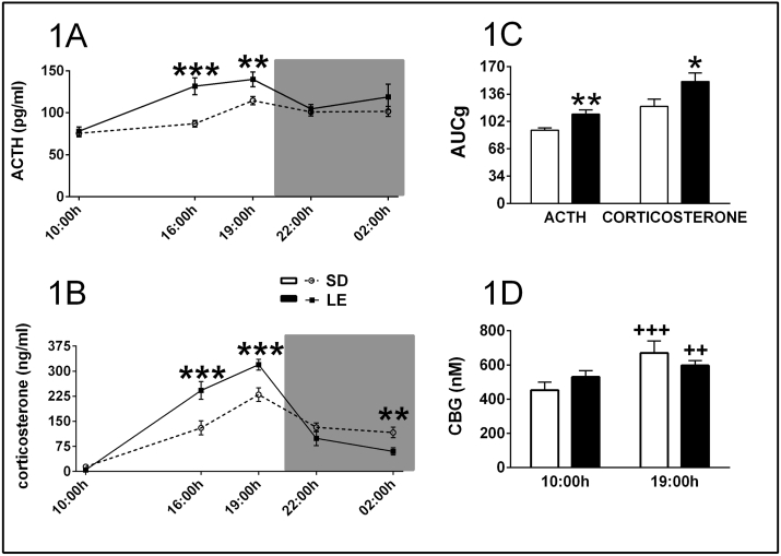
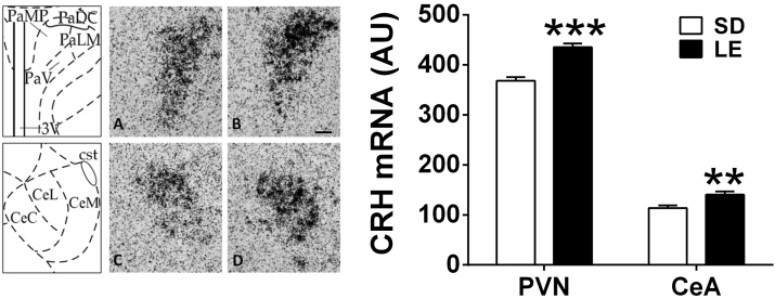
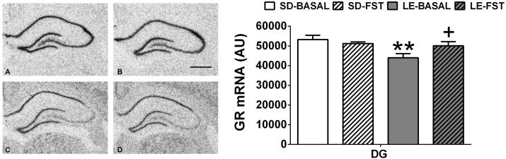
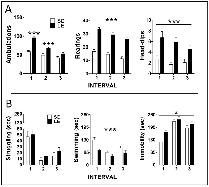
Rat and mouse strains differ in behavioral and physiological characteristics, and such differences can contribute to explain discrepant results between laboratories and better select the most appropriate strain for a particular purpose. Differences in the activity of the hypothalamic-pituitary-adrenal (HPA) axis are particularly important given the pivotal role of this system in determining consequences of exposure to stressors. In this regard, Long-Evans (LE) rats are widely used in stress research, but there is no specific study aiming at thoroughly characterizing HPA activity in LE versus other extensively used strains. In a first experiment, LE showed higher resting ACTH and corticosterone levels only at certain points of the circadian rhythm, but much greater ACTH responsiveness to stressors (novel environment and forced swim) than Sprague-Dawley (SD) rats. Accordingly, enhanced corticotropin-releasing hormone (CRH) expression in the paraventricular nucleus of the hypothalamus and reduced expression of glucocorticoid receptors were observed in the hippocampal formation. Additionally, they are hyperactive in novel environments, and prone to adopt passive-like behavior when compared to SD rats. Supporting that altered HPA function has a marked physiological impact, we observed in another set of animals much lower thymus weight in LE than SD rats. Finally, to demonstrate that LE rats are likely to have higher HPA responsiveness to stressors than most strains, we studied resting and stress levels of HPA hormones in LE versus Wistar and Fischer rats, the latter considered an example of high HPA responsiveness. Again, LE showed higher resting and stress levels of ACTH than both Wistar and Fischer rats. As ACTH responsiveness to stressors in LE rats is stronger than that previously reported when comparing other rat strains and they are commercially available, they could be an appropriate model for studying the behavioral and physiological implications of a hyper-active HPA axis under normal and pathological conditions.

Keywords: Corticosteroid receptors; Corticotropin-releasing hormone; Hypothalamic-pituitary-adrenal axis; Long-Evans; Strain differences; Stress responsiveness.


Conflict of interest statement

The authors have no conflicts of interest to declare.

Figures

Fig. 1
Fig. 1
LE and SD differences in the basal levels of peripheral pituitary-adrenal variables. It is represented Mean ± SEM (n = 14 per group) of the circadian pattern of plasma levels of ACTH and corticosterone (A and B), the area under the curve (AUCg) of the two hormones over the 24 h period (Panel C), and the plasma levels of CBG at two time points; ***p ≤ 0.001, **p ≤ 0.01, *p ≤ 0.05 vs SD; +++ p ≤ 0.001, ++ p ≤ 0.01 vs. respective 10:00 h sampling time.
Fig. 2
Fig. 2
LE and SD differences in the ACTH and corticosterone response to two acute stressors (15 min): hole-board (HB) and forced swim test. Mean ± SEM are represented (n = 14 for the HB, n = 8 for the forced swim test). Basal levels obtained on a different day were similar in the two strains and are represented with dotted lines. Blood sampling was done between 09.00 and 13.00 h. Hormone levels were higher in LE than SD rats except for corticosterone after the forced swim test; ***p ≤ 0.001, **p ≤ 0.01 vs SD.
Fig. 3
Fig. 3
LE and SD differences in CRH mRNA levels in the PVN and the CeA. Mean ± SEM of arbitrary units are represented (n = 13–14 per group). Brains were processed under non-stress conditions or immediately after the forced swim test. Since there were no differences between non-stress and stress conditions, all animals from the same strain were represented and analyzed together. Expression was studied in the areas showed in the left panels, corresponding to the mpdPVN (PaMP) and the lateral CeA (CeL). A and B: representative images of the PVN in SD (left) and LE (right) rats. C and D: representative images of CeA in SD (left) and LE (right) (100 μm scale bar); ***p ≤ 0.001, **p ≤ 0.01 vs SD.
Fig. 4
Fig. 4
LE and SD differences in GR mRNA levels in the dentate gyrus (DG). Mean ± SEM of arbitrary units are represented (n = 5–6 for basal conditions; n = 7–8 for FST stress). A and B: representative images of MR mRNA expression in the hippocampal formation in SD (left) and LE (right) rats. C and D: representative images of GR mRNA expression SD (left) and LE (right). 500 μm scale bar. GR mRNA expression was lower in LE strain at basal levels in comparison to SD rats; **p ≤ 0.01 vs. SD-Basal, + p ≤ 0.05 vs. LE-Basal.
Fig. 5
Fig. 5
Behavior of LE and SD rats during hole-board (HB) and forced swim exposure (15 min). Mean ± SEM of different behaviors (are represented in 5 min blocks (upper panel: HB; lower panel: forced swim). Number of rats per group were 14 in the HB and 8 in the forced swim test (the remaining rats were perfused under resting conditions). LE rats showed higher levels of ambulation during the first 10 min, and overall higher levels of rearing and head-dips at all times. ***p ≤ 0.01 vs SD strain. During forced swim, there were overall lower levels of swimming and higher levels of immobility in LE vs SD rats (strain effect); ***p ≤ 0.001, *p ≤ 0.05.
Fig. 6
Fig. 6
Resting and stress levels of HPA hormones in Wistar, Fischer and LE rats. Means ± SEM (n = 10) are represented. Please note the different scales for both hormones depending on the experimental condition (panel A: resting levels at lights on and at the peak around lights off; Panel B and C: responses to hole-board and forced swim exposure for 15 min). * Indicates differences versus Wistar, + differences versus Fischer (one symbol p ≤ 0.05; two symbols p ≤ 0.01; three symbols p ≤ 0.001.

References

    1. Abel E.L. Response to alarm substance in different rat strains. Physiol. Behav. 1992;51:345–347. - PubMed
    1. Akana S.F., Cascio C.S., Shinsako J., Dallman M.F. Corticosterone: narrow range required for normal body and thymus weight and ACTH. Am. J. Physiol. 1985;249:527–532. - PubMed
    1. Altemus M., Smith M.A., Diep V., Aulakh C.S., Murphy D.L. Increased mRNA for corticotrophin releasing hormone in the amygdala of fawn-hooded rats: a potential animal model of anxiety. Anxiety. 1994-1995;1:251. 157. - PubMed
    1. Armario A., Gavaldà A., Martí O. Forced swimming test in rats: effect of desipramine administration and the period of exposure to the test on struggling behavior, swimming, immobility and defecation rate. Eur. J. Pharmacol. 1988;158:207–212. - PubMed
    1. Armario A., Nadal R. Individual differences and the characterization of animal models of psychopathology: a strong challenge and a good opportunity. Front. Pharmacol. 2013;4:137. - PMC - PubMed