

Primary orbital ganglioneuroblastoma: A case report
- PMID: 34307890
- PMCID: PMC8284332
- DOI: 10.1515/med-2021-0230
Primary orbital ganglioneuroblastoma: A case report
Abstract
Background: Ganglioneuroblastoma (GNB) is a neoplasm that arises from the primitive cells of the sympathetic nervous system during childhood. The current case is very unique because of the initial primary tumor manifestation in the orbit and an adrenal tumor being observed later during follow-up.
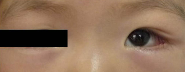
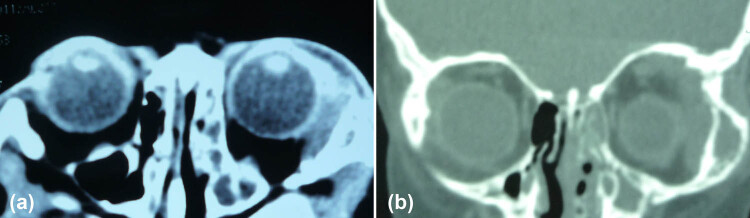
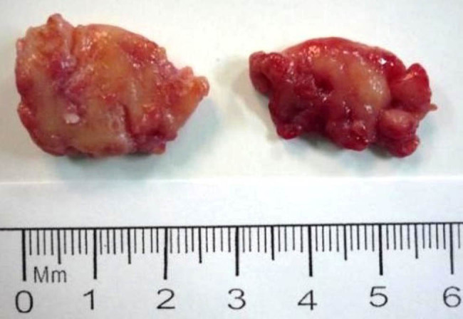
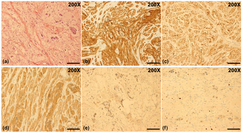
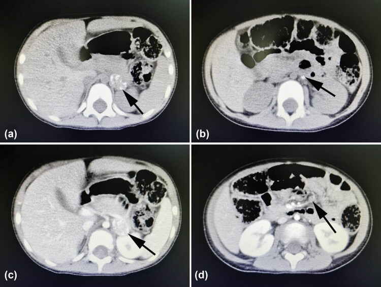
Case presentation: A 2-year-old girl presented to the Ophthalmology Department of our hospital complaining of swelling of the left upper eyelid for approximately one month. Orbital computed tomography (CT) revealed a left orbital mass with bone destruction. Thoracic and abdominal CT indicated no abnormalities. The mass was surgically resected, and histopathological analysis confirmed it as GNB. During follow-up, abdominal CT detected an adrenal tumor with internal calcification, a calcified nodule on the left side of the abdominal aorta, and mesenteric lymph nodes. Accordingly, primary orbital GNB and metastatic adrenal GNB were the possible considerations. We removed the adrenal tumor, and the patient underwent chemotherapy. However, the patient died 18 months after the ophthalmic surgery.
Conclusion: Primary orbital GNB in children is easily misdiagnosed because of its rare occurrence and atypical clinical findings. Imaging methods combined with histopathological examination contribute to the detection and diagnosis of primary and metastatic GNBs. Thus, timely surgery combined with adjuvant chemotherapy and long-term follow-up is essential for controlling the metastasis of GNB and improving the survival rate of patients.
Keywords: adrenal gland; orbit; primary ganglioneuroblastoma.
© 2021 Ruixin Ma et al., published by De Gruyter.
Conflict of interest statement
Conflict of interest: None of the authors reports conflicts of interest in this work.
Figures
Similar articles
-
Primary orbital ganglioneuroblastoma in a child: A case report.Medicine (Baltimore). 2020 Nov 6;99(45):e22922. doi: 10.1097/MD.0000000000022922. Medicine (Baltimore). 2020. PMID: 33157934 Free PMC article.
-
Adult-onset adrenal ganglioneuroblastoma - Bone metastasis two years after surgery: report of a case.Surg Today. 2010 May;40(5):482-6. doi: 10.1007/s00595-008-4084-0. Epub 2010 Apr 28. Surg Today. 2010. PMID: 20425556
-
Review of our experience with neuroblastoma and ganglioneuroblastoma in adults.World J Surg. 2014 Nov;38(11):2871-4. doi: 10.1007/s00268-014-2682-0. World J Surg. 2014. PMID: 25002244
-
Primary intracranial pediatric ganglioneuroblastoma-report of two cases and review of an unusual masquerader.Childs Nerv Syst. 2024 Dec;40(12):4289-4294. doi: 10.1007/s00381-024-06578-3. Epub 2024 Aug 24. Childs Nerv Syst. 2024. PMID: 39180696 Review.
-
[A case of composite pheochromocytoma-ganglioneuroblastoma in the adrenal gland with primary hyperparathyroidism].Hinyokika Kiyo. 2003 May;49(5):269-72. Hinyokika Kiyo. 2003. PMID: 12822455 Review. Japanese.
Cited by
-
Stereotactic radiosurgery for brain metastases secondary to adrenal ganglioneuroblastoma: illustrative case.J Neurosurg Case Lessons. 2025 Mar 24;9(12):CASE24820. doi: 10.3171/CASE24820. Print 2025 Mar 24. J Neurosurg Case Lessons. 2025. PMID: 40127476 Free PMC article.
-
Nonspecific Gastrointestinal Symptoms as the First Sign of Ganglioneuroblastoma Intermixed-Case Report and Literature Review.J Clin Med. 2023 Sep 21;12(18):6092. doi: 10.3390/jcm12186092. J Clin Med. 2023. PMID: 37763032 Free PMC article. Review.
References
-
- Youlden DR, Jones BC, Cundy TP, Karpelowsky J, Aitken JF, McBride CA. Incidence and outcomes of neuroblastoma in Australian children: a population-based study (1983–2015). J Paediat Child Health 2020;56(7):1046–52. - PubMed
- Youlden DR, Jones BC, Cundy TP, Karpelowsky J, Aitken JF, McBride CA. Incidence and outcomes of neuroblastoma in Australian children: a population-based study (1983–2015) J Paediat Child Health. 2020;56(7):1046–52. - PubMed
-
- Shimada H, Ambros IM, Dehner LP, Hata JI, Joshi VV, Roald B, et al. The international neuroblastoma pathology classification (the Shimada system). Cancer. 1999;86(2):364–72. - PubMed
- Shimada H, Ambros IM, Dehner LP, Hata JI, Joshi VV, Roald B. et al. The international neuroblastoma pathology classification (the Shimada system) Cancer. 1999;86(2):364–72. - PubMed
-
- Wahl HR, Craig PE. Report of a case showing a distinct ganglioneuroma, a neroblastoma and a cystic calcifying ganglioneuroblastoma. Am J Pathol. 1938;14:797–808. - PMC - PubMed
- Wahl HR, Craig PE. Report of a case showing a distinct ganglioneuroma, a neroblastoma and a cystic calcifying ganglioneuroblastoma. Am J Pathol. 1938;14:797–808. - PMC - PubMed
-
- Emre Ş, Zcan R, Bakır AC, Kuruğoğlu S, Omunoğlu N, Şen HS, et al. Adrenal masses in children: imaging, surgical treatment and outcome. Asian J Surg. 2020;43(1):207–12. - PubMed
- Emre Ş, Zcan R, Bakır AC, Kuruğoğlu S, Omunoğlu N, Şen HS. et al. Adrenal masses in children: imaging, surgical treatment and outcome. Asian J Surg. 2020;43(1):207–12. - PubMed
-
- Salas M, Esparza H. A case of ganglioneuroblastoma with metastasis to the orbital bones. Boletin Med del Hospital Infant de Mexico. 1963;20:343–60. - PubMed
- Salas M, Esparza H. A case of ganglioneuroblastoma with metastasis to the orbital bones. Boletin Med del Hospital Infant de Mexico. 1963;20:343–60. - PubMed
Publication types
LinkOut - more resources
Full Text Sources