

Oropharyngeal primary tumor segmentation for radiotherapy planning on magnetic resonance imaging using deep learning
- PMID: 34307917
- PMCID: PMC8295848
- DOI: 10.1016/j.phro.2021.06.005
Oropharyngeal primary tumor segmentation for radiotherapy planning on magnetic resonance imaging using deep learning
Abstract
Background and purpose: Segmentation of oropharyngeal squamous cell carcinoma (OPSCC) is needed for radiotherapy planning. We aimed to segment the primary tumor for OPSCC on MRI using convolutional neural networks (CNNs). We investigated the effect of multiple MRI sequences as input and we proposed a semi-automatic approach for tumor segmentation that is expected to save time in the clinic.
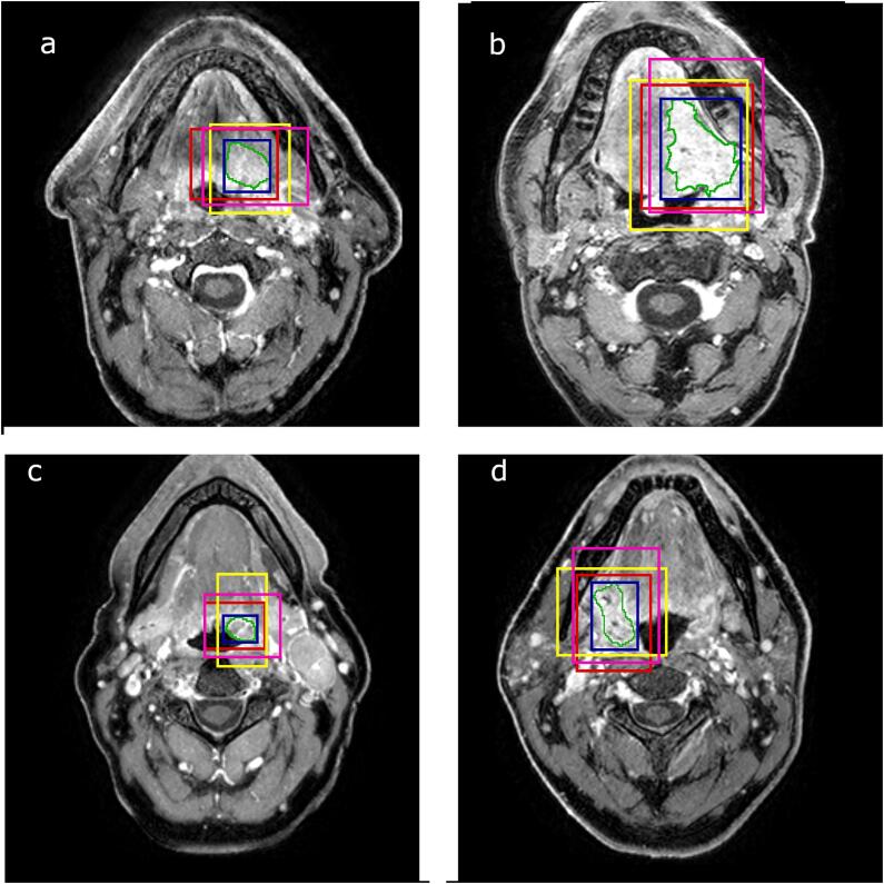
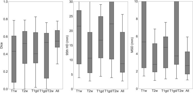
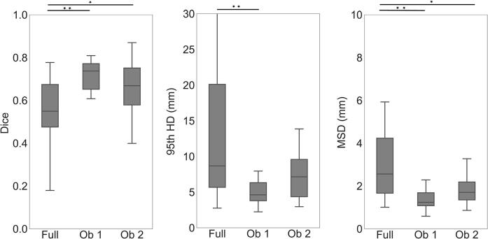
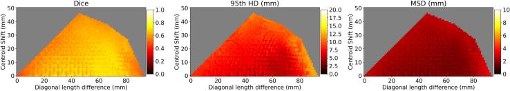
Materials and methods: We included 171 OPSCC patients retrospectively from 2010 until 2015. For all patients the following MRI sequences were available: T1-weighted, T2-weighted and 3D T1-weighted after gadolinium injection. We trained a 3D UNet using the entire images and images with reduced context, considering only information within clipboxes around the tumor. We compared the performance using different combinations of MRI sequences as input. Finally, a semi-automatic approach by two human observers defining clipboxes around the tumor was tested. Segmentation performance was measured with Sørensen-Dice coefficient (Dice), 95th Hausdorff distance (HD) and Mean Surface Distance (MSD).
Results: The 3D UNet trained with full context and all sequences as input yielded a median Dice of 0.55, HD of 8.7 mm and MSD of 2.7 mm. Combining all MRI sequences was better than using single sequences. The semi-automatic approach with all sequences as input yielded significantly better performance (p < 0.001): a median Dice of 0.74, HD of 4.6 mm and MSD of 1.2 mm.
Conclusion: Reducing the amount of context around the tumor and combining multiple MRI sequences improved the segmentation performance. A semi-automatic approach was accurate and clinically feasible.
Keywords: Convolutional neural network; MRI; Oropharyngeal cancer; Radiotherapy; Segmentation; Semi-automatic approach.
© 2021 The Authors. Published by Elsevier B.V. on behalf of European Society of Radiotherapy & Oncology.
Conflict of interest statement
The authors declare that they have no known competing financial interests or personal relationships that could have appeared to influence the work reported in this paper.
Figures

References
-
- Fitzmaurice C., Allen C., Barber R.M., Barregard L., Bhutta Z.A., Brenner H. Global, regional, and national cancer incidence, mortality, years of life lost, years lived with disability, and disability-adjusted life-years for 32 cancer groups, 1990 to 2015. JAMA Oncol [Internet] 2017;3:524. doi: 10.1001/jamaoncol.2016.5688. - DOI - PMC - PubMed
-
- D Souza AM, Chen L, Wu Y, Abidin AZ, Xu C, Wismüller A. MRI tumor segmentation with densely connected 3D CNN. 2018. SPIE Medical Imaging Proceedings, 10574. doi: 10.1117/12.2293394.
-
- Li J., Chen H., Li Y., Peng Y. ACM international conference proceeding series. 2019. A novel network based on densely connected fully convolutional networks for segmentation of lung tumors on multi-modal MR images; pp. 1–5. doi: 10.1145/3358331.3358400.
LinkOut - more resources
Full Text Sources
