. 2021 Jun 25:11:100070.
doi: 10.1016/j.nbscr.2021.100070. eCollection 2021 Nov.

Chronic methamphetamine uncovers a circadian rhythm in multiple-unit neural activity in the dorsal striatum which is independent of the suprachiasmatic nucleus

Affiliations

Chronic methamphetamine uncovers a circadian rhythm in multiple-unit neural activity in the dorsal striatum which is independent of the suprachiasmatic nucleus

Shota Miyazaki et al. Neurobiol Sleep Circadian Rhythms. .

Abstract

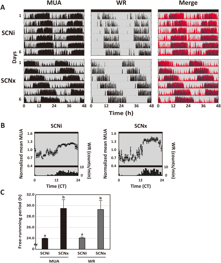
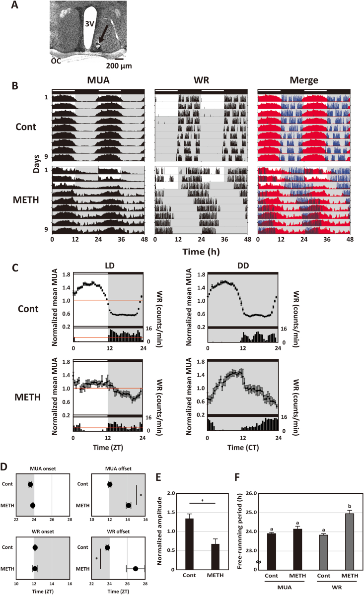
The dorsal striatum forms part of the basal ganglia circuit that is a major regulator of voluntary motor behavior. Dysfunction in this circuit is a critical factor in the pathology of neurological (Parkinson's and Huntington's disease) as well as psychiatric disorders. In this study, we employed in vivo real-time monitoring of multiple unit neural activity (MUA) in the dorsal striatum of freely moving mice. We demonstrate that the striatum exhibits robust diurnal and circadian rhythms in MUA that peak in the night. These rhythms are dependent upon the central circadian clock located in the suprachiasmatic nucleus (SCN) as lesions of this structure caused the loss of rhythmicity measured in the striatum. Nonetheless, chronic treatment of methamphetamine (METH) makes circadian rhythms appear in MUA recorded from the striatum of SCN-lesioned mice. These data demonstrate that the physiological properties of neurons in the dorsal striatum are regulated by the circadian system and that METH drives circadian rhythms in striatal physiology in the absence of the SCN. The finding of SCN-driven circadian rhythms in striatal physiology has important implications for an understanding of the temporal regulation of motor control as well as revealing how disease processes may disrupt this regulation.

Keywords: Circadian rhythm; Dopamine; Methamphetamine; Multiple unit neural activity; Striatum; Suprachiasmatic nucleus; Wheel running activity; constant dark, (DD); dopamine transporter, (DAT); dopamine, (DA); light-dark, (LD); methamphetamine, (METH); multiple unit neural activity, (MUA); suprachiasmatic nucleus, (SCN); wheel running, (WR).


Conflict of interest statement

None.

Figures

Fig. 1
Fig. 1
Simultaneous recording of MUA in the striatum and WR rhythms in freely moving SCNi and SCNx mice. (A) A typical histological section indicates the recording site with a neutral red stain. The black arrowhead indicates the recording site in the striatum. The scale bar is 300 μm. (B) Representative double-plotted actograms showing diurnal and circadian rhythms of MUA in the striatum, WR and merged in SCNi mice. The red and blue actograms indicate MUA and WR, respectively. The mice were maintained in the LD cycle and then transferred into the DD condition. Lighting conditions are indicated at the top of the figure; open bars indicate a light phase and closed indicate a dark phase. (C) Representative integrated mean activities of MUA and WR in SCNi mice were plotted according to LD cycle and DD condition. MUA in the LD cycle for 3 days was normalized to 24 h in 30 min bins; each point indicates a level of MUA counts at the time relative to average MUA counts at 24 h. WR in the LD cycle for 3 days was plotted in 60 min bins. Red line indicates the standard mean activity of MUA and WR. As in the LD cycle, each MUA and WR in the DD condition for 6 days were also plotted to CT. Data were shown as means ± SEM in 30 min bins (MUA) or 60 min bins (WR). (D) The activity onsets and offsets of MUA in the striatum and WR in SCNi mice were calculated under the LD cycle. (E) The free-running periods of MUA in the striatum and WR in SCNi mice were calculated. *p < 0.05, Student's unpaired t-test. (F) A typical histological section indicates that the SCN was lesioned by a positive current. The scale bar is 200 μm. (G) Representative double-plotted actograms showing the diurnal and circadian rhythms of MUA in the striatum, WR, and merged in SCNx mice. The red and blue actograms indicate MUA and WR, respectively. The mice were maintained in the LD cycle and then transferred into the DD condition. The lighting conditions are indicated at the top of the figure; open bars indicate light phase and closed indicate dark. (H) Representative integrated mean activities of MUA and WR in SCNx mice plotted in LD and DD. Each MUA and WR in LD for 3 days were normalized to 24 h. Each MUA and WR in DD for 6 days were normalized to 24 h. . (For interpretation of the references to colour in this figure legend, the reader is referred to the Web version of this article.)
Fig. 2
Fig. 2
In vivo MUA rhythms in the striatum of METH-treated SCNi and SCNx mice. (A) Representative double-plotted actograms showing circadian rhythms of MUA in the striatum, WR and merged in METH-treated SCNi and SCNx mice. The red and blue actograms indicate MUA and WR, respectively. The actograms represent DD conditions for 6 days. (B) Representative integrated mean activities of MUA and WR plotted under the DD condition. MUA in the DD condition for 6 days was normalized to a circadian time in 30 min bins; each point indicates a level of MUA counts at the time relative to average MUA counts at a circadian time. WR in the DD condition for 6 days was plotted in 60 min bins to a circadian time. Data were shown as means ± SEM in 30 min bins (MUA) or 60 min bins (WR). (C) The free-running periods of MUA in the striatum and WR in METH-treated SCNi and SCNx mice were calculated. Differing letters between groups indicate significant differences (p < 0.05, Tukey's test). (For interpretation of the references to colour in this figure legend, the reader is referred to the Web version of this article.)
Fig. 3
Fig. 3
In vivo MUA rhythms in the SCN of METH-treated mice. (A) A typical histological section indicates the recording site using a neutral red stain. The black arrowhead indicates the recording site in the SCN. The scale bar indicates 200 μm. (B) Representative actograms showing diurnal and circadian rhythms of MUA in the SCN, WR and merged in METH-untreated (Cont) and METH-treated (METH) mice. The red and blue actograms indicate MUA and WR, respectively. The mice were maintained in the LD cycle and then transferred into the DD condition. The lighting conditions are indicated at the top of the figure; open bars indicate light phase and closed indicate dark. (C) Representative integrated mean activities of MUA and WR in Cont and METH-treated mice were plotted according to LD cycle and DD condition. MUA in the LD cycle for 3 days was normalized to 24 h in 30 min bins; each point indicates a level of MUA counts at the time relative to average MUA counts at 24 h. WR in the LD cycle for 3 days was plotted in 60 min bins. Red line indicates the standard mean activity of MUA and WR. As in the LD cycle, each MUA and WR in the DD condition for 6 days were also plotted to CT. Data were shown as means ± SEM in 30 min bins (MUA) or 60 min bins (WR). (D) The activity onsets and offsets of MUA in the SCN and WR in Cont and METH-treated mice were calculated in the LD cycle, respectively. *p < 0.05, Student's unpaired t-test. (E) The amplitude of MUA in the SCN in Cont and METH-treated mice were calculated in the LD cycle. *p < 0.05, Student's unpaired t-test. (F) The free-running periods of MUA in the SCN and WR in Cont and METH-treated mice were calculated. Differing letters between groups indicate significant differences (p < 0.05, Tukey's test). (For interpretation of the references to colour in this figure legend, the reader is referred to the Web version of this article.)

References

    1. Blum I.D., Zhu L., Moquin L., Kokoeva M.V., Gratton A., Giros B., Storch K.F. A highly tunable dopaminergic oscillator generates ultradian rhythms of behavioral arousal. Elife. 2014;3 doi: 10.7554/eLife.05105. - DOI - PMC - PubMed
    1. Brami-Cherrier K., Lewis R.G., Cervantes M., Liu Y., Tognini P., Baldi P., Sassone-Corsi P., Borrelli E. Cocaine-mediated circadian reprogramming in the striatum through dopamine D2R and PPARγ activation. Nat. Commun. 2020;11:4448. doi: 10.1038/s41467-020-18200-6. - DOI - PMC - PubMed
    1. De Corte B.J., Wagner L.M., Matell M.S., Narayanan N.S. Striatal dopamine and the temporal control of behavior. Behav. Brain Res. 2019;356:375–379. doi: 10.1016/j.bbr.2018.08.030. - DOI - PMC - PubMed
    1. Ferris M.J., España R.A., Locke J.L., Konstantopoulos J.K., Rose J.H., Chen R., Jones S.R. Dopamine transporters govern diurnal variation in extracellular dopamine tone. Proc. Natl. Acad. Sci. U.S.A. 2014;111:E2751–E2759. doi: 10.1073/pnas.1407935111. - DOI - PMC - PubMed
    1. Grippo R.M., Purohit A.M., Zhang Q., Zweifel L.S., Güler A.D. Direct midbrain dopamine input to the suprachiasmatic nucleus accelerates circadian entrainment. Curr. Biol. 2017;27:2465–2475. doi: 10.1016/j.cub.2017.06.084. - DOI - PMC - PubMed