Review
. 2021 Aug 4;22(8):e52507.
doi: 10.15252/embr.202152507. Epub 2021 Jul 26.

Maintaining proteostasis under mechanical stress

Affiliations
Review

Maintaining proteostasis under mechanical stress

Jörg Höhfeld et al. EMBO Rep. .

Abstract

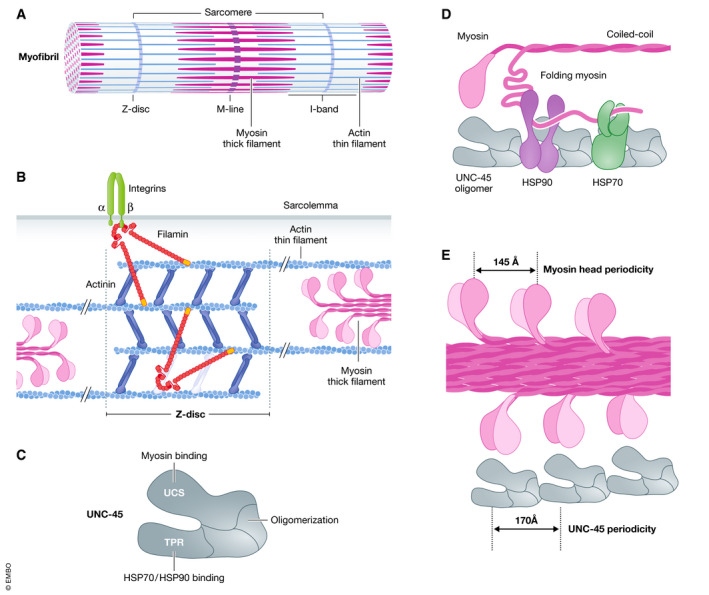
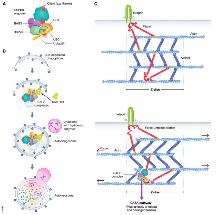
Cell survival, tissue integrity and organismal health depend on the ability to maintain functional protein networks even under conditions that threaten protein integrity. Protection against such stress conditions involves the adaptation of folding and degradation machineries, which help to preserve the protein network by facilitating the refolding or disposal of damaged proteins. In multicellular organisms, cells are permanently exposed to stress resulting from mechanical forces. Yet, for long time mechanical stress was not recognized as a primary stressor that perturbs protein structure and threatens proteome integrity. The identification and characterization of protein folding and degradation systems, which handle force-unfolded proteins, marks a turning point in this regard. It has become apparent that mechanical stress protection operates during cell differentiation, adhesion and migration and is essential for maintaining tissues such as skeletal muscle, heart and kidney as well as the immune system. Here, we provide an overview of recent advances in our understanding of mechanical stress protection.

Keywords: autophagy; chaperones; mechanobiology; proteostasis; signal transduction.


Conflict of interest statement

The authors declare that they have no conflict of interest.

Figures

Figure 1
Figure 1. Protecting the proteome against mechanical stress relies on protein folding and degradation systems that recognize force‐unfolded proteins
(A) Stress protection systems comprise chaperone/cochaperone complexes and specialized E2/E3 ubiquitin conjugation systems, which recognize unfolded proteins under stress conditions to facilitate their refolding or their degradation by the proteasome and autophagic/lysosomal pathways. Ubiquitylation can be assisted by molecular chaperones or can proceed through a direct recognition of unfolded proteins by quality control E3 ubiquitin ligases. Stress‐induced signalling pathways regulate the activity of the involved folding and degradation systems. (B) Cellular systems are permanently exposed to a wide variety of mechanical signals. After recognition and transmission through cell–cell and cell–matrix contacts as well as cytoskeletal systems, these signals induce a variety of specific cell responses. (C) Mechanical forces can trigger the unfolding of mechanosensory proteins such as talin, which links integrin‐containing adhesion complexes in the plasma membrane (PM) to the actin cytoskeleton (ECM—extracellular matrix).
Figure 2
Figure 2. The UNC‐45‐containing chaperone system mediates the folding and assembly of myosin in muscle sarcomeres
(A) The sarcomere represents the smallest contractile unit of striated muscles. It is repetitively arranged in tubular myofibrils, numerous bundles of which form the muscle fibre. Z‐discs limit the sarcomere on both sides and mediate the anchoring of actin thin filaments. Myosin thick filaments are intercalated between the actin filaments and are connected at the M‐line. The I‐band is the region that contains exclusively actin filaments. (B) Actin and filamin crosslink actin filaments within the Z‐disc. In addition, filamin interacts with integrin molecules in the sarcolemma. (C) Schematic representation of the domain structure of the cochaperone UNC‐45. UCS, UNC‐45/CRO1/She4p domain and TPR, tetratricopeptide repeat. (D) Oligomeric UNC‐45 coordinates the activity of HSP70 and HSP90 chaperone proteins during the folding and assembly of myosin filaments. (E) The UNC‐45 oligomer provides a molecular scaffold for enforcing the regular spacing of folded myosin head domains in the myosin thick filament.
Figure 3
Figure 3. The BAG3 chaperone complex initiates a force‐induced autophagy pathway in contracting muscle
(A) BAG3 links chaperones of the HSP70 family to small heat shock proteins such as HSPB8. The HSP70‐associated ubiquitin ligase CHIP can associate with the BAG3 chaperone complex to ubiquitylate a bound client protein in cooperation with a ubiquitin conjugation enzyme (UBC). (B) Enclosure of the BAG3 chaperone complex by phagophore membranes is facilitated by the autophagic ubiquitin adaptor SQSTM1, leading to autophagosome formation and ultimately to degradation in autolysosomes. (C) Actin–myosin contraction causes the force‐induced unfolding of the actin‐crosslinking protein filamin at the Z‐disc of the muscle sarcomere. Mechanically unfolded and damaged forms of filamin are recognized by the BAG3 complex and directed onto the CASA pathway for disposal.
Figure 4
Figure 4. Podocytes withstand high mechanical forces at the kidney filtration barrier
(A) Schematic representation of the kidney glomerulus. Blood is filtered across the walls of the glomerular capillaries lined with podocytes. The filtrate enters the urinary space and exits through the proximal tubule. (B) The glomerular filtration barrier is formed by a fenestrated endothelium, the glomerular basement membrane and podocytes. Podocytes possess long foot processes that wrap around the capillaries, leaving slit diaphragms (Sd) available for blood filtration. (C) At the slit diaphragm, foot processes are connected by a mechanosensitive protein complex containing nephrin, Neph1 and podocin. The complex is linked to TRPC6 ion channels, diverse kinases and the actin cytoskeleton. Filamin mediates actin anchoring and crosslinking at the slit diaphragm.
Figure 5
Figure 5. Mechanical forces govern the adhesion, migration and activity of immune cells
(A) Immune cells, such as leukocytes, are subjected to variety of mechanical forces when they leave the blood stream and migrate through sites of infection and inflammation. (B) Schematic representation of the integrin machinery that operates in immune cells. The transduction of mechanical signals between intracellular processes and the extracellular matrix (ECM) via integrin heterodimers requires a two‐way communication system that employs cytoskeletal adapters (e.g. talin, kindlin, vinculin), kinases (e.g. FAK, Src) and mediators of Rho‐GTPase signalling. Integrin engagement with extracellular ligands affects motility and cell fate decisions, including survival, proliferation and differentiation via downstream signalling cascades. PM, plasma membrane.

References

    1. Adriaenssens E, Geuens T, Baets J, Echaniz‐Laguna A, Timmerman V (2017) Novel insights in the disease biology of mutant small heat shock proteins in neuromuscular diseases. Brain 140: 2541–2549 - PubMed
    1. Alon R, Dustin ML (2007) Force as a facilitator of integrin conformational changes during leukocyte arrest on blood vessels and antigen‐presenting cells. Immunity 26: 17–27 - PubMed
    1. Alon R, Ley K (2008) Cells on the run: shear‐regulated integrin activation in leukocyte rolling and arrest on endothelial cells. Curr Opin Cell Biol 20: 525–532 - PMC - PubMed
    1. Altmann SM, Grünberg RG, Lenne P‐F, Ylänne J, Raae A, Herbert K, Saraste M, Nilges M, Hörber JKH (2002) Pathways and intermediates in forced unfolding of spectrin repeats. Structure 10: 1085–1096 - PubMed
    1. Arimura T, Ishikawa T, Nunoda S, Kawai S, Kimura A (2011) Dilated cardiomyopathy‐associated BAG3 mutations impair Z‐disc assembly and enhance sensitivity to apoptosis in cardiomyocytes. Hum Mutat 32: 1481–1491 - PubMed

Publication types