. 2021 Jul;9(14):e14979.
doi: 10.14814/phy2.14979.

The impact of hindlimb disuse on sepsis-induced myopathy in mice

Affiliations

The impact of hindlimb disuse on sepsis-induced myopathy in mice

Orlando Laitano et al. Physiol Rep. 2021 Jul.

Abstract

Sepsis induces a myopathy characterized by loss of muscle mass and weakness. Septic patients undergo prolonged periods of limb muscle disuse due to bed rest. The contribution of limb muscle disuse to the myopathy phenotype remains poorly described. To characterize sepsis-induced myopathy with hindlimb disuse, we combined the classic sepsis model via cecal ligation and puncture (CLP) with the disuse model of hindlimb suspension (HLS) in mice. Male C57bl/6j mice underwent CLP or SHAM surgeries. Four days after surgeries, mice underwent HLS or normal ambulation (NA) for 7 days. Soleus (SOL) and extensor digitorum longus (EDL) were dissected for in vitro muscle mechanics, morphological, and histological assessments. In SOL muscles, both CLP+NA and SHAM+HLS conditions elicited ~20% reduction in specific force (p < 0.05). When combined, CLP+HLS elicited ~35% decrease in specific force (p < 0.05). Loss of maximal specific force (~8%) was evident in EDL muscles only in CLP+HLS mice (p < 0.05). CLP+HLS reduced muscle fiber cross-sectional area (CSA) and mass in SOL (p < 0.05). In EDL muscles, CLP+HLS decreased absolute mass to a smaller extent (p < 0.05) with no changes in CSA. Immunohistochemistry revealed substantial myeloid cell infiltration (CD68+) in SOL, but not in EDL muscles, of CLP+HLS mice (p < 0.05). Combining CLP with HLS is a feasible model to study sepsis-induced myopathy in mice. Hindlimb disuse combined with sepsis induced muscle dysfunction and immune cell infiltration in a muscle dependent manner. These findings highlight the importance of rehabilitative interventions in septic hosts to prevent muscle disuse and help attenuate the myopathy.

Keywords: atrophy; infection; inflammation; muscle; septic shock; weakness.


Conflict of interest statement

Authors declare that they have no conflict of interest to disclose.

Figures

FIGURE 1
FIGURE 1
Combination of cecal ligation and puncture (CLP) with hindlimb suspension (HLS) as a pre‐clinical model to study sepsis‐induced skeletal muscle abnormalities. Mice were suspended 4 days after CLP surgeries. HLS lasted for 7 days and muscle tissue was collected for functional and morphological assessment. SOL = soleus, EDL = extensor digitorum longus
FIGURE 2
FIGURE 2
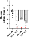
Changes in body mass induced by each experimental intervention. # < 0.05 in comparison to Sham+NA group. Data are mean and standard error (SE). Two‐way ANOVA with multiple comparisons
FIGURE 3
FIGURE 3
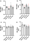
Muscle mass for SOL (panels a & b) and EDL (panels c & d) normalized by body mass (mg/g) and expressed as absolute (mg). Data are mean and standard error (SE). Two‐way ANOVA with multiple comparisons. # different from Sham+NA; * different from CLP+NA
FIGURE 4
FIGURE 4
Median fiber areas show that CLP+HLS resulted in atrophy in SOL (panel a), but not in EDL muscles (panel c). Data are median and standard error (SE). Two‐way ANOVA with multiple comparisons. Cumulative percentage fiber size distributions highlight the leftward shift of the fiber area distribution, indicating that a greater proportion of fibers were smaller (panels b and d). # different from Sham+NA
FIGURE 5
FIGURE 5
Soleus (SOL) (panel a) force‐frequency curves. Lines represent best fit using sigmoidal Hill equation. SOL maximal absolute (panel b) and specific forces (panel c); peak twitch (panel d) and half‐relaxation time (Panel e). SOL maximal specific force was markedly reduced in SHAM+HLS and CLP+NA mice and further reduced in CLP+HLS mice. Peak twitch was reduced in SHAM+HLS, CLP+NA, and CLP+HLS mice. # different from Sham+NA; * different from CLP+NA
FIGURE 6
FIGURE 6
Extensor digitorum longus (EDL) (panel a) force‐frequency curves. Lines represent best fit using sigmoidal Hill equation. EDL maximal absolute (panel b) and specific (panel c) forces; peak twitch (panel d) and half‐relaxation time (panel e). EDL force was only reduced by the combination of CLP+HLS. Data are mean and standard error (SE). Two‐way ANOVA with multiple comparisons. # different from Sham+NA; * different from CLP+NA; & different from Sham+HLS
FIGURE 7
FIGURE 7
Increased infiltration of CD68+ cells was observed in SOL muscle (panel a) in the CLP+HLS group. No changes were observed in EDL muscles (panel b). Data are mean and standard error (SE). Two‐way ANOVA with multiple comparisons. Representative sections of SOL and EDL depicting the greater accumulation of macrophages (in green) in CLP+HLS condition for SOL muscles. White squares at the top panels represent areas of magnification in the bottom panels. # different from Sham+NA; * different from CLP+NA

References

    1. Augusto, V. , Padovani, C. R. , & Campos, G. E. R. (2004). Skeletal muscule fiber types in C57BL6J mice. Brazilian Journal of Morphological Science 21, 89–94.
    1. Brakenridge, S. C. , Efron, P. A. , Stortz, J. A. , Ozrazgat‐Baslanti, T. , Ghita, G. , Wang, Z. , Bihorac, A. , Mohr, A. M. , Brumback, B. A. , Moldawer, L. L. , & Moore, F. A. (2018). The impact of age on the innate immune response and outcomes after severe sepsis/septic shock in trauma and surgical intensive care unit patients. Journal of Trauma and Acute Care Surgery, 85, 247–255. 10.1097/TA.0000000000001921 - DOI - PMC - PubMed
    1. Callahan, L. A. , & Supinski, G. S. (2009). Sepsis‐induced myopathy. Critical Care Medicine, 37, S354–S367. 10.1097/CCM.0b013e3181b6e439 - DOI - PMC - PubMed
    1. Chleboun, G. S. , Patel, T. J. , & Lieber, R. L. (1997). Skeletal muscle architecture and fiber‐type distribution with the multiple bellies of the mouse extensor digitorum longus muscle. Acta Anatomica, 159, 147–155. 10.1159/000147977 - DOI - PubMed
    1. Côt́e, C. H. , Tremblay, M.‐H. , Duchesne, E. , & Lapoite, B. M. (2008). Inflammation‐induced leukocyte accumulation in injured skeletal muscle: Role of mast cells. Muscle & Nerve, 37, 754–763. 10.1002/mus.20998 - DOI - PubMed

Publication types