. 2022 Feb;28(3-4):150-160.
doi: 10.1089/ten.TEA.2021.0057. Epub 2021 Oct 7.

Limited Expansion of Human Hepatocytes in FAH/ RAG2-Deficient Swine

Affiliations

Limited Expansion of Human Hepatocytes in FAH/ RAG2-Deficient Swine

Erek David Nelson et al. Tissue Eng Part A. 2022 Feb.

Abstract

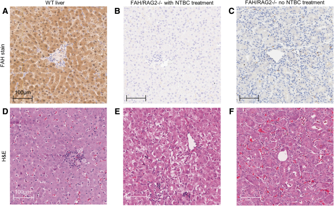
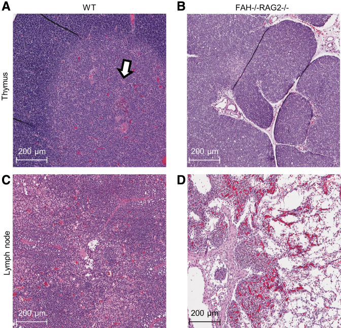
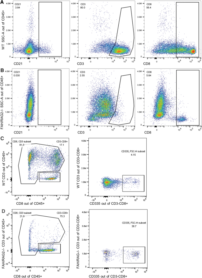
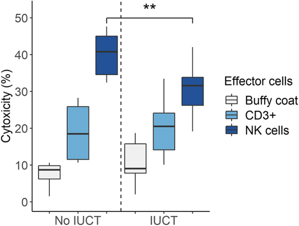
The mammalian liver's regenerative ability has led researchers to engineer animals as incubators for expansion of human hepatocytes. The expansion properties of human hepatocytes in immunodeficient mice are well known. However, little has been reported about larger animals that are more scalable and practical for clinical purposes. Therefore, we engineered immunodeficient swine to support expansion of human hepatocytes and identify barriers to their clinical application. Immunodeficient swine were engineered by knockout of the recombinase-activating gene 2 (RAG2) and fumarylacetoacetate hydrolase (FAH). Immature human hepatocytes (ihHCs) were injected into fetal swine by intrauterine cell transplantation (IUCT) at day 40 of gestation. Human albumin was measured as a marker of engraftment. Cytotoxicity against ihHCs was measured in transplanted piglets and control swine. We initially detected higher levels of human albumin in cord blood of newborn FAH/RAG2-deficient (FR) pigs compared with immunocompetent controls (196.26 ng/dL vs. 39.29 ng/dL, p = 0.008), indicating successful engraftment of ihHCs after IUCT and adaptive immunity in the fetus. Although rare hepatocytes staining positive for human albumin were observed, levels of human albumin did not rise after birth, but declined, suggesting rejection of xenografted ihHCs. Cytotoxicity against ihHCs increased after birth by 3.8% (95% CI: [2.1%-5.4%], p < 0.001) and inversely correlated with declining levels of human albumin (p = 2.1 × 10⁻⁵, R2 = 0.17). Circulating numbers of T cells and B cells were negligible in FR pigs. However, circulating natural killer (NK) cells exerted cytotoxicity against ihHCs. NK cell activity was lower in immunodeficient piglets after IUCT than in naive controls (30.4% vs. 40.1%, p = 0.011, 95% CI for difference [2.7%-16.7%]). In conclusion, ihHCs were successfully engrafted in FR swine after IUCT. NK cells were a significant barrier to expansion of hepatocytes. New approaches are needed to overcome this hurdle and allow large-scale expansion of human hepatocytes in immunodeficient swine. Impact statement There is currently a need for robust expansion of human hepatocytes. We describe an immunodeficient swine model into which we engrafted immature human hepatocytes (ihHCs). We identified the mechanism of the eventual graft rejection by the intact NK cell population, which has not been previously shown to have a significant role in xenograft rejection. By both improving engraftment and reducing NK cell-mediated cytotoxicity toward the graft through intrauterine cell transfer, we confirmed the presence of residual adaptive immunity in this model of immunodeficiency and the ability to induce hyposensitization in the NK cell population by taking advantage of the fetal microenvironment.

Keywords: animal model; genetic immunodeficiency; hepatocyte transplant; hereditary tyrosinemia; natural killer cell.


Conflict of interest statement

S.N., R.H., and J.L. are inventors of FAH-deficient swine. The FAH-deficient swine patent is owned by Mayo Clinic and Oregon Health and Science University and licensed to Cytotheryx (Rochester, MN). J.L. is a board member of Cytotheryx.

Figures

FIG. 1.
FIG. 1.
Immunohistochemical and H&E staining of liver tissue of FR pigs demonstrates no visible FAH+ hepatocyte xenograft and no inflammatory infiltrate or vasculopathy indicative of acute rejection. (A) WT tissue with FAH staining showing diffuse cytoplasmic expression. (B) FR pigs on NTBC, with FAH staining. (C) FR pigs on no NTBC treatment, with FAH staining. (B, C) Loss of FAH expression without visible FAH+ cells. (D) WT tissue with H&E staining. (E) FR pig on NTBC, with H&E staining. Hepatocellular ballooning is evident in zone 3. (F) FR pig on no NTBC treatment, with H&E staining, demonstrates dysplastic change with atypical hepatocyte architecture and pseudoacinar forms. Neither (E) nor (F) demonstrates vasculopathy or lymphocytic, neutrophilic, or macrocytic infiltrate, which would be evident with acute xenograft rejection. H&E, hematoxylin and eosin.
FIG. 2.
FIG. 2.
H&E staining demonstrates a markedly abnormal structure of lymphoid organs in FR pigs consistent with the immunodeficient phenotype. (A) WT thymus with normal architecture and expected corticomedullary differentiation. Hassall's corpuscles (arrow) are seen. (B) FR pig thymus. FR pig sample demonstrates hypoplasia, reduction in lobule size, and lack of corticomedullary differentiation. (C) WT lymph node. (D) FR pig lymph node, with depleted lymphocyte population that has been replaced with fibrofatty tissue. Lymph node architecture is preserved.
FIG. 3.
FIG. 3.
Peripheral blood leukocytes of FR pigs demonstrate reduced T cell and B cell populations, but normal levels of NK cells. (A) WT pig serum forward and side scatter area distributions of CD21, CD3, and CD8 of CD45+ cells, nonspecific lymphocyte markers. (B) FR pig serum demonstrates significant absence of CD21+, decreased CD3+, and decreased CD8+ cells, consistent with the relative paucity of T cells and B cells in the RAG2−/− phenotype. (C) WT pig serum CD3 and CD8 sorting, then further analysis of the CD335 population of the CD3CD8+ subset. (D) FR pig serum CD3 and CD8 sorting, demonstrating a CD3CD8+ population that is not depleted when compared with the WT animal. Further gating of CD335+ and CD3CD8+ demonstrates persistent populations of both CD335+ and CD335 cells.
FIG. 4.
FIG. 4.
Serum human albumin production by transplanted human hepatocytes in FR piglets is increased at birth compared with immunocompetent piglets, while hepatocytes staining positive for human albumin are observed. Human albumin production subsequently shows a downtrend. (A) Serum human albumin at birth, measured in 50 piglets. FR pigs had significantly higher levels of human albumin at birth than control Fah−/− pigs (p = 0.008, Mann–Whitney U test). The addition of RAG2−/− increased engraftment of functional human hepatocytes in the porcine liver. (B) Serum human albumin in four FR piglets measured sequentially after birth. Pig A also received a portal vein injection of ihHCs on day 43 postbirth, indicated by (*) in the image, and serum human albumin concentration was measured on days 2, 10, and 27 after this injection. Human albumin levels declined across all time points in all piglets. (C) Representative anti-human albumin IHC slide of FR pig liver. Rare hepatocytes staining positive for human albumin (white arrows) are scattered throughout the livers of FR pigs who underwent IUCT with ihHCs on gestational day 40. No associated inflammatory cells are present. IUCT, intrauterine cell transplantation; ihHC, immature human hepatocyte.
FIG. 5.
FIG. 5.
Concentration-dependent NK cell cytotoxicity was reduced after IUCT. PBLs of three FR pigs were tested for cytotoxic activity against ihHCs biweekly from 6 to 14 weeks of age; two pigs were post-IUCT and one pig was not post-IUCT. Cytotoxicity increased with further purification of NK cells. NK cell-mediated cytotoxicity was reduced in two FR pigs after IUCT compared with the non-IUCT FR pig (**p = 0.011, t-test). Cytotoxicity of the buffy coat and CD3+ cells was similar in all FR pigs. Of note, no significant trends in NK cell cytotoxicity were observed over time within an individual FR pig. PBL, peripheral blood leukocyte.

References

    1. Matsunari, H., Nagashima, H., Watanabe, M., et al. . Blastocyst complementation generates exogenic pancreas in vivo in apancreatic cloned pigs. PNAS 110, 4557, 2013. - PMC - PubMed
    1. Kobayashi, T., Yamaguchi, T., Hamanaka, S., et al. . Generation of rat pancreas in mouse by interspecific blastocyst injection of pluripotent stem cells. Cell 142, 787, 2010. - PubMed
    1. Ogle, B.M., Butters, K.A., Plummer, T.B., et al. . Spontaneous fusion of cells between species yields transdifferentiation and retroviral transfer in vivo. FASEB J 18, 548, 2004. - PubMed
    1. Ogle, B.M., Cascalho, M., and Platt, J.L.. Biological implications of cell fusion. Nature reviews. Mol Cell Biol 6, 567, 2005. - PubMed
    1. Grompe, M. Fah knockout animals as models for therapeutic liver repopulation. Adv Exp Med Biol 959, 215, 2017. - PubMed

Publication types

LinkOut - more resources