

Know thy immune self and non-self: Proteomics informs on the expanse of self and non-self, and how and where they arise
- PMID: 34310018
- PMCID: PMC8865197
- DOI: 10.1002/pmic.202000143
Know thy immune self and non-self: Proteomics informs on the expanse of self and non-self, and how and where they arise
Abstract
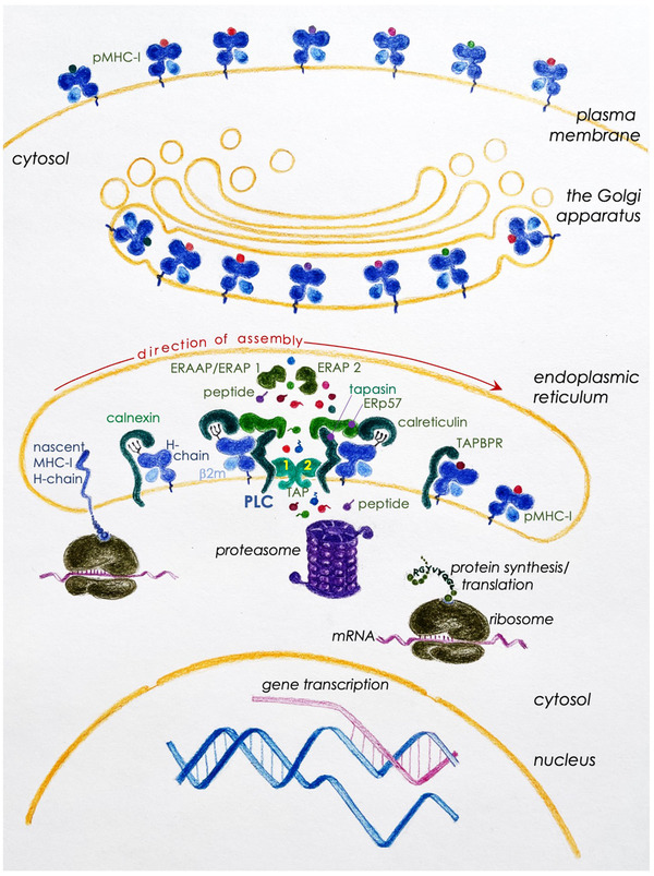
T cells play an important role in the adaptive immune response to a variety of infections and cancers. Initiation of a T cell mediated immune response requires antigen recognition in a process termed MHC (major histocompatibility complex) restri ction. A T cell antigen is a composite structure made up of a peptide fragment bound within the antigen-binding groove of an MHC-encoded class I or class II molecule. Insight into the precise composition and biology of self and non-self immunopeptidomes is essential to harness T cell mediated immunity to prevent, treat, or cure infectious diseases and cancers. T cell antigen discovery is an arduous task! The pioneering work in the early 1990s has made large-scale T cell antigen discovery possible. Thus, advancements in mass spectrometry coupled with proteomics and genomics technologies make possible T cell antigen discovery with ease, accuracy, and sensitivity. Yet we have only begun to understand the breadth and the depth of self and non-self immunopeptidomes because the molecular biology of the cell continues to surprise us with new secrets directly related to the source, and the processing and presentation of MHC ligands. Focused on MHC class I molecules, this review, therefore, provides a brief historic account of T cell antigen discovery and, against a backdrop of key advances in molecular cell biologic processes, elaborates on how proteogenomics approaches have revolutionised the field.
Keywords: T cell epitope; antigen presentation; antigen processing; human leukocyte antigen; immunopeptidomics; major histocompatibility complex; mass spectrometry.
© 2021 The Authors. Proteomics published by Wiley-VCH GmbH.
Conflict of interest statement
The authors declare no conflict of interest.
Figures
Similar articles
-
Discrimination Between Human Leukocyte Antigen Class I-Bound and Co-Purified HIV-Derived Peptides in Immunopeptidomics Workflows.Front Immunol. 2018 Apr 27;9:912. doi: 10.3389/fimmu.2018.00912. eCollection 2018. Front Immunol. 2018. PMID: 29780384 Free PMC article.
-
Determination of a Predictive Cleavage Motif for Eluted Major Histocompatibility Complex Class II Ligands.Front Immunol. 2018 Aug 6;9:1795. doi: 10.3389/fimmu.2018.01795. eCollection 2018. Front Immunol. 2018. PMID: 30127785 Free PMC article.
-
NNAlign_MA; MHC Peptidome Deconvolution for Accurate MHC Binding Motif Characterization and Improved T-cell Epitope Predictions.Mol Cell Proteomics. 2019 Dec;18(12):2459-2477. doi: 10.1074/mcp.TIR119.001658. Epub 2019 Oct 2. Mol Cell Proteomics. 2019. PMID: 31578220 Free PMC article.
-
[MHC tetramers: tracking specific immunity].Acta Med Croatica. 2003;57(4):255-9. Acta Med Croatica. 2003. PMID: 14639858 Review. Croatian.
-
Antigen presenting cells.Immunol Res. 1989;8(2):98-117. doi: 10.1007/BF02919073. Immunol Res. 1989. PMID: 2659691 Review.
Cited by
-
From immune equilibrium to immunodynamics.Front Microbiol. 2022 Nov 25;13:1018817. doi: 10.3389/fmicb.2022.1018817. eCollection 2022. Front Microbiol. 2022. PMID: 36504800 Free PMC article.
-
Unlocking the secrets of the immunopeptidome: MHC molecules, ncRNA peptides, and vesicles in immune response.Front Immunol. 2025 Jan 29;16:1540431. doi: 10.3389/fimmu.2025.1540431. eCollection 2025. Front Immunol. 2025. PMID: 39944685 Free PMC article. Review.
-
East meets west: integrating Yin-Yang theory with immunology teaching.Front Immunol. 2024 Aug 20;15:1441863. doi: 10.3389/fimmu.2024.1441863. eCollection 2024. Front Immunol. 2024. PMID: 39229266 Free PMC article. Review.
-
The Importance of Being Presented: Target Validation by Immunopeptidomics for Epitope-Specific Immunotherapies.Front Immunol. 2022 Apr 6;13:883989. doi: 10.3389/fimmu.2022.883989. eCollection 2022. Front Immunol. 2022. PMID: 35464395 Free PMC article. Review.
References
-
- Davis, D. (2014). The compatibility gene: How our bodies fight disease, attract others, and define ourselves, Oxford University Press.
-
- Snell, G. D. , Dausset, J. , & Nathenson, S. G. (1976). Histocompatibility, Academic Press.
-
- Zinkernagel, R. M. , & Doherty, P. C. (1974). Immunological surveillance against altered self components by sensitised T lymphocytes in lymphocytic choriomeningitis. Nature, 251(5475), 547–548. - PubMed
-
- Zinkernagel, R. M. , & Doherty, P. C. (1974). Restriction of in vitro T cell‐mediated cytotoxicity in lymphocytic choriomeningitis within a syngeneic or semiallogeneic system. Nature, 248(5450), 701–702. - PubMed
-
- Klein, J. (1994). Alone on the heart of the earth: An immunogeneticist's journey into the past. Advances in Cancer Research, 63, 1–39. - PubMed
Publication types
MeSH terms
Substances
Grants and funding
- N01 AI040079/AI/NIAID NIH HHS/United States
- I01 BX001444/BX/BLRD VA/United States
- IK6 BX004595/BX/BLRD VA/United States
- R01 DE027749/DE/NIDCR NIH HHS/United States
- A21988/CRUK_/Cancer Research UK/United Kingdom
- R01 HL054977/HL/NHLBI NIH HHS/United States
- R21 AI042284/AI/NIAID NIH HHS/United States
- R01 HL121139/HL/NHLBI NIH HHS/United States
- R01 AI042284/AI/NIAID NIH HHS/United States
- R01 AI137082/AI/NIAID NIH HHS/United States
- A28736/CRUK_/Cancer Research UK/United Kingdom
- I01 BX006010/BX/BLRD VA/United States
LinkOut - more resources
Full Text Sources
Research Materials
