. 2021 Jul 26;17(7):e1008603.
doi: 10.1371/journal.ppat.1008603. eCollection 2021 Jul.

DENV NS1 and MMP-9 cooperate to induce vascular leakage by altering endothelial cell adhesion and tight junction

Affiliations

DENV NS1 and MMP-9 cooperate to induce vascular leakage by altering endothelial cell adhesion and tight junction

Pan Pan et al. PLoS Pathog. .

Abstract

Dengue virus (DENV) is a mosquito-borne pathogen that causes a spectrum of diseases including life-threatening dengue hemorrhagic fever (DHF) and dengue shock syndrome (DSS). Vascular leakage is a common clinical crisis in DHF/DSS patients and highly associated with increased endothelial permeability. The presence of vascular leakage causes hypotension, circulatory failure, and disseminated intravascular coagulation as the disease progresses of DHF/DSS patients, which can lead to the death of patients. However, the mechanisms by which DENV infection caused the vascular leakage are not fully understood. This study reveals a distinct mechanism by which DENV induces endothelial permeability and vascular leakage in human endothelial cells and mice tissues. We initially show that DENV2 promotes the matrix metalloproteinase-9 (MMP-9) expression and secretion in DHF patients' sera, peripheral blood mononuclear cells (PBMCs), and macrophages. This study further reveals that DENV non-structural protein 1 (NS1) induces MMP-9 expression through activating the nuclear factor κB (NF-κB) signaling pathway. Additionally, NS1 facilitates the MMP-9 enzymatic activity, which alters the adhesion and tight junction and vascular leakage in human endothelial cells and mouse tissues. Moreover, NS1 recruits MMP-9 to interact with β-catenin and Zona occludens protein-1/2 (ZO-1 and ZO-2) and to degrade the important adhesion and tight junction proteins, thereby inducing endothelial hyperpermeability and vascular leakage in human endothelial cells and mouse tissues. Thus, we reveal that DENV NS1 and MMP-9 cooperatively induce vascular leakage by impairing endothelial cell adhesion and tight junction, and suggest that MMP-9 may serve as a potential target for the treatment of hypovolemia in DSS/DHF patients.


Conflict of interest statement

The authors have declared that no competing interests exist.

Figures

Fig 1
Fig 1. DENV induces the production of NS1 and MMP-9 in severe dengue patients.
(A and B) The serum concentrations of NS1(A) and MMP-9 (B) in healthy donors and severe dengue patients infected days (2, 5, 8, and 11 days) were measured by ELISA. Points represent the value in each serum sample. (C and D) The serum concentrations of NS1 (C) and MMP-9 (D) in each severe dengue patients infected days (2, 5, 8, and 11 days) were measured by ELISA. Points represent the value in each serum sample. (E) the correlations of the concentrations of NS1 and MMP-9 in the same group of severe dengue patients infected 11 days were plotted. Linear regressions were traced according to the distributions of the points. Dates were representative of two independent experiments. ns means not significant. P ≤ 0.05 (*), P ≤ 0.01 (**), P ≤ 0.001 (***).
Fig 2
Fig 2. DENV NS1 interacts with MMP-9.
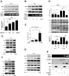
(A) HEK293T cells were co-transfected with HA-MMP-9 and Flag-Cap, Flag-M, Flag-Prm, Flag-E, Flag-NS1, Flag-NS2A, Flag-NS2B, Flag-NS3, Flag-NS4A, or Flag-NS4B. Cell lysates were immunoprecipitataed using anti-Flag antibody, and analyzed using anti-Flag and anti-HA antibody. Cell lysates (40 μg) was used as Input. (B) HEK293T cells were co-transfected with HA-MMP9 and Flag-NS1, Cell lysates were immunoprecipitataed using anti-HA antibody, and analyzed using anti-Flag and anti-HA antibody. Cell lysates (40 μg) was used as Input. (C, D) HEK293T cells were co-transfected with empty vector or VC-155-MMP-9 and VN173-NS1/E/NS4A. At 24 h post-transfection, living cells were observed by confocal microscopy. The quantification of YFP-positive cells was determined by ImageJ software (D). ND means not detection. (E) Purified His-SARS-CoV-2-N (5 μg) or His-DENV2-NS1 (5 μg) was incubated with purified no-tagged MMP-9 protein (3 μg) for 24 h, Mixtures were incubated with Ni-NTA Agarose beads. Mixtures were analyzed by immunoblotting using anti-MMP9, anti-NS1, anti-His antibody. Untreated protein including His-SARS-CoV-2-N (1 μg), His-DENV2-NS1 (1 μg), or no-tagged MMP-9 protein (1 μg) were analyzed by immunoblotting using anti-MMP9, anti-NS1, and anti-His antibody (as input). (F) Yeast strain AH109 were co-transformed with combination of binding domain (BD-p53, BD-MMP-9, and BD-Lam) and activation domain (AD-T, AD-NS1) plasmid. Transfected yeast cells were grown on SD-minus Trp/Leu double dropout plates, and colonies were replicated on to SD-minus Trp/Leu/Ade/His fourth dropout plates to check for the expression of reporter genes. (G, H) Schematic diagram of wild-type MMP-9 protein and truncated mutants MMP-9 protein (D1 to D9) (G). HEK293T cells were co-transfected with HA-NS1 and Flag-MMP-9 truncated mutants (D1 to D9). Cell lysates were immunoprecipitated using anti-Flag antibody, and analyzed using anti-Flag and anti-HA antibody. Cell lysates (40 μg) was used as Input (H). Dates were representative of three independent experiments.
Fig 3
Fig 3. NS1 induces expression and proteolytic activity of MMP-9.
(A) PMA-differentiated THP-1 macrophages were transfected with the different concentrations of plasmid encoding NS1 for 24 h. Cell lysates were analyzed (top) by immunoblotting. Supernatants were analyzed (middle) by gelatin zymography assays for MMP-9 proteinase activity. Intracellular MMP-9 RNA (bottom) was determined by qRT-PCR analysis. (B) HEK293T cells were co-transfected with the plasmid encoding MMP-9 and different concentrations of plasmid encoding NS1 for 24 h. Cell lysates were analyzed (top) by immunoblotting. Supernatants were analyzed (middle) by gelatin zymography assays for MMP-9 proteinase activity. Intracellular MMP-9 RNA (bottom) was determined by qRT-PCR analysis. (C) HEK293T cells were con-transfected with different concentrations of NS1 expressing plasmid and NF-κB reporter plasmid. Luciferase assays were performed 20 h after transfection. (D) PMA-differentiated THP-1 macrophages were firstly transfected with plasmid encoding HA-CT or HA-NS1 for 20 h, and then treated with 200 nM SC75741 for 5 h, MMP-9 protein in cell supernatants were measured by ELISA (top) and indicated proteins in cell extract were analyzed by WB (middle). Intracellular MMP-9 RNA (bottom) was determined by qRT-PCR analysis. (EG) Hela cells (E), HEK293T cells (F), and PMA-differentiated THP-1 macrophages (G) were transfected with different concentrations of plasmid encoding NS1 for 24 h. The indicated proteins in cell extract were analyzed by WB. (H) The supernatants of HUVEC cells were incubated with BSA (3 μg/ml), NS1 (3 μg/ml), pro-MMP-9 protein (200 ng/ml), BSA (3 μg/ml) plus pro-MMP-9 protein (200 ng/ml), NS1 (3 μg/ml) plus pro-MMP-9 protein (200 ng/ml) or MMP-9 protein (200 ng/ml) for 6 h, and then the supernatants were analyzed by gelatin zymography assays for MMP-9 proteinase activity and the indicated protein expression were analyzed by immunoblotting. Dates were representative of two to three independent experiments. ns means not significant. Values are mean ± SEM, P ≤ 0.05 (*), P ≤ 0.01 (**), P ≤ 0.001 (***).
Fig 4
Fig 4. DENV2 induces MMP-9 expression and secretion in human PBMCs and macrophages, but not in HUVECs.
(A) PMA-differentiated THP-1 macrophages (top) or HUVEC cells (bottom) were treated with infectious or UV- inactivated DENV2 at MOI = 5 for 48h. Intracellular DENV2 E RNA (bottom) was determined by qRT-PCR analysis. Mock: untreated cells. Control: supernatant of C6/36 cells without DENV2 infection. (B and C) PMA-differentiated THP-1 macrophages were infected with DENV2 for different times at MOI = 5 (B) or at different MOI for 24 h (C). Intracellular MMP-9 RNA (top) and DENV2 E RNA (bottom) was determined by qRT-PCR analysis, MMP-9 proteinase activity in the supernatants was determined by gelatin zymography assays and proteins in cell extract (middle) were analyzed by Western blotting. (D and E) HUVEC cells were infected with DENV2 for different times at MOI = 5 (DC) and at different MOI for 24 h (E). Intracellular MMP-9 RNA (top) and DENV2 E RNA (bottom) was determined by qRT-PCR analysis, MMP-9 proteinase activity in the supernatants was determined by gelatin zymography assays and proteins in cell extract (middle) were analyzed by Western blotting. (F) HUVEC cells or PMA-differentiated THP-1 macrophages were infected with DENV2 at MOI = 5 for 24 h. Viral copies were quantified by RT-PCR. (G) HUVEC cells or PMA-differentiated THP-1 macrophages were equally distributed to four 12-hole plates and infected with DENV2 at MOI = 5 for 24 h. MMP-9 protein in cell supernatants were measured by ELISA (top) and indicated proteins in cell extract were analyzed by WB (bottom). Dates were representative of three independent experiments. ns means not significant. Values are mean ± SEM, P ≤0.05 (*), P ≤0.01 (**), P ≤0.001 (***).
Fig 5
Fig 5. NS1 facilitates MMP-9 to induce endothelial hyperpermeability in human cells and mice tissues.
(A) PMA-differentiated THP-1 macrophages were infected with DENV2 for different times at MOI = 5, NS1 protein in Supernatants were analyzed by ELISA (top). Cell lysates were analyzed by immunoblotting (bottom). (B) Confluent monolayers of HUVEC cells were grown on polycarbonate membrane system and treated with the supernatants came from DENV2 infected HUVEC cells or THP-1 cells for 24 h or pre-incubated with 600nM SB-3CT (a specific inhibitor of MMP-9 protein) or 600 nM SC75741 for 1h. Endothelial permeability was evaluated by measuring trans-endothelial electrical resistance (TEER) (ohm) using EVOM2 epithelial voltohmmeter. (CI) IFNAR-/- C57BL/6 mice were intravenously injected with 300 μl DENV2 at a dose of 1×10⁶ PFU/mouse (n = 6), pre-treated with 300 μl PBS containing MMP-9 specific inhibitor SB-3CT (5 mg/kg per mice) by intraperitoneal injection for 90 min and then treated with DENV2 (1×10⁶ PFU/mouse), repeat treated with SB-3CT (5 mg/kg per mice) on the fourth day after DENV2 (NGC) infection (n = 6), or 300 μl PBS containing the same volume DMSO as a control group (n = 4). 7 days after infection, mice were euthanasia, and the tissues were collected. MMP-9 RNA in the blood was determined by qRT-PCR (upper) and MMP-9 protein in the serum was measured by ELISA (lower). Points represent the value of each serum samples (C). Evans blue dye was intravenously injected into mice 7 days after DENV infected groups (n = 5), control groups (n = 4) and DENV+SB-3CT (n = 5) (C–E). The dye was allowed to circulate for 2 hours before mice were euthanasia, tissues include liver (D), spleen (E) and lung (F) were collected, and the value of Evans blue was measured at OD610. Histopathology analysis of tissues includes Liver (G), Spleen (H) and Lung (I) after DENV infection. (J) Monolayers of HUVEC cells grown on Transwell inserts were incubated for 48 h with MMP-9 protein (100 ng/ml) or NS1 protein (5 μg/ml) or NS1 (5 μg/ml) plus different concentration of MMP-9 (50 ng/ml to 100ng/ml) or pre-treated with 600 nM SB-3CT or 600 nM SC7574 for 1 h, then incubated with NS1 plus MMP-9. The TEER (ohm) was measured at indicated time points. Dates were representative of two to three independent experiments. ns means not significant. Values are mean ± SEM, P ≤0.05 (*), P ≤0.01 (**), P ≤0.001 (***).
Fig 6
Fig 6. NS1 recruits MMP-9 to disrupts the junctions between endothelial cells.
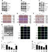
(AF) IFNAR-/- C57BL/6 mice were intravenously injected with 300 μl DENV2 at a dose of 1×10⁶ PFU/mouse (n = 6), pre-treated with 300 μl PBS containing MMP-9 specific inhibitor SB-3CT (5 mg/kg per mice) by intraperitoneal injection for 90 min and then treated with DENV2 (1×10⁶ PFU/mouse), repeat treated with SB-3CT (5 mg/kg per mice) on the fourth day after DENV2 (NGC) infection (n = 6), or 300 μl PBS containing the same volume DMSO as a control group (n = 4). 7 days after infection, mice were euthanasia, and the tissues were collected. The indicated proteins in Lung (A), spleen (B) and Liver (C) were measured by Western-blot. The expression of β-catenin in Liver (D), spleen (E), and Lung (F) by Immunohistochemistry analysis. (G) HUVEC cells were respectively transfected with plasmid encoding MMP-9 (2 μg) or NS1 (2 μg) or NS1 (1 ug) plus MMP-9 (1 μg) for 24 h or firstly co-transfected with plasmid encoding NS1 (1ug) plus MMP-9 (1 μg) for 12 h, then treated with 600nM SB-3CT for 12 h. The indicated proteins in cell extract were analyzed by WB. (H–K) HUVEC cells were treated with NS1 protein (5 μg/ml) or MMP-9 protein (100 ng/ml) or NS1 (5 μg/ml) plus MMP-9 (100 ng/ml) or pre-incubated with 600 nM SB-3CT for 1 h, then treated with NS1 (5 μg/ml) plus MMP-9 (100 ng/ml) for 6 h, The distribution of endogenous β-catenin (H) or ZO-1 (J) protein were visualized under confocal microscope. The quantifications of relative β-catenin (I) or ZO-1 (K) intensities were determined by ImageJ software. The quantification of protein was used by ImageJ software (A–G). (D–F) +++ means Percentage contribution of high positive cells; ++ means Percentage contribution of positive cells; + means Percentage contribution of low positive cells;—means Percentage contribution of negative cells. ns means not significant. All dates were representative of two to three independent experiments.
Fig 7
Fig 7. NS1 recruits MMP-9 to interact with adhesion and tight junction proteins.
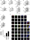
(A) HEK293T cells were transfected with plasmid encoding HA-NS1 plus Flag-β-catenin. Cell lysates were immunoprecipitated using anti-HA antibody, and analyzed using anti-Flag, anti-HA, or anti-β-catenin antibody. Cell lysates (40 μg) were used as Inputs. (B) Hela cells were transfected with plasmid encoding HA-NS1, Cell lysates were immunoprecipitated using anti-HA antibody, and analyzed using anti-HA or anti-β-catenin antibody. Cell lysates (40 μg) was used as Input. (C, D) HEK293T cells (C) or Hela cells (D) were co-transfected with plasmid encoding HA-NS1 plus Flag-ZO-1, Cell lysates were immunoprecipitated using anti-HA antibody, and analyzed using anti-Flag or anti-HA antibody. Cell lysates (40 μg) were used as Inputs. (E, F) HEK293T cells were transfected with Flag-β-catenin (E) or Flag-ZO-1 (F) and incubated with purified His-SARS-CoV-2-N (5 μg) or His-DENV2-NS1 (5 μg) for 24 h, cell extracts were incubated with Ni-NTA Agarose beads. Mixtures were analyzed by immunoblotting using anti-β-catenin, anti-NS1, anti-His, anti-ZO-1 antibody. Untreated proteins including His-SARS-CoV-2-N (1 μg) and His-DENV2-NS1 (1 μg) and HEK293T cell lysates were analyzed by immunoblotting using anti-β-catenin, anti-NS1, anti-His, and anti-ZO-1 antibody (as input). (G, H) Hela cells were co-transfected with plasmid encoding HA-NS1 plus Flag-MMP-9, Cell lysates were immunoprecipitated using anti-Flag (G) or anti-β-catenin antibody (H), and analyzed using anti-Flag, anti-HA or anti-β-catenin antibody. Cell lysates (40 μg) was used as Input. (I, J) Hela cells were co-transfected with plasmid encoding HA-NS1, HA-MMP-9, and Flag-ZO-1 (I) or Flag-NS1, HA-MMP-9, and Flag-ZO-1 (J). Cell lysates were immunoprecipitated using anti-Flag (top) or anti-HA antibody (bottom), and analyzed using anti-Flag, anti-HA or anti-MMP-9 antibody. Cell lysates (40 μg) was used as Input. (K, L) HUVEC cells were treated with NS1 protein (5 μg/ml), MMP-9 protein (100 ng/ml), and NS1 protein (5 μg/ml) plus MMP-9 protein (100 ng/ml), respectively, for 6 h. The distributions of the membrane marker (Dil) (yellow), the endogenous β-catenin (yellow) and ZO-1 (yellow) proteins and the extracellular NS1 (red) and MMP-9 (green) proteins were visualized under confocal microscope. The quantifications of co-localization fluorescence were determined by ImageJ software (L). ND means not detected. All dates were representative of three independent experiments.
Fig 8
Fig 8. NS1 induce vascular leakage through recruiting MMP-9 in mice.
C57BL/6 mice and MMP-9-/- mice were injected intravenously DENV2 NS1 protein [10 mg/kg (n = 5)], the same volume of PBS was also tail vein injected to C57BL/6 mice and MMP-9-/- mice (n = 5) as control group. Another group of MMP-9-/- mice (n = 5) were injected intravenously DENV2 NS1 protein (10 mg/kg) plus recombinant mouse MMP-9 protein (70 μg /kg). (AC) After 24 h post-injection, mice were intravenously injected with Evans blue dye. The dye was allowed to circulate for 2h before mice were euthanized, and tissue include Lung (A), spleen (B), and Liver (C) were collected. The value of Evans blue was measured at OD610. (DI) After 24 h post-injection, mice were euthanized and tissue were collected. The indicated proteins in Lung (D), spleen (E), and Liver (F) were measured by Western-blot. The expression of β-catenin in Lung (G), spleen (H), and Liver (I) were analyzed by Immunohistochemistry. All dates were representative of two to three independent experiments. The quantification of protein was used by ImageJ software (D–I). (G–H) +++ means Percentage contribution of high positive cells; ++ means Percentage contribution of positive cells; + means Percentage contribution of low positive cells;—means Percentage contribution of negative cells. ns means not significant. Values are mean ± SEM, P ≤0.05 (*), P ≤0.01 (**), P ≤0.001 (***).
Fig 9
Fig 9. A proposed model in which DENV NS1 and MMP-9 coordinate to induce vascular leakage by altering endothelial cell adhesion and tight junctions.
DENV non-structural protein 1 (NS1) induces MMP-9 expression through activating the nuclear factor κB (NF-κB) signaling pathway. Additionally, NS1 interacts with MMP-9 and facilitates the enzyme to alter the adhesion and tight junctions and vascular leakage in human endothelial cells and mice tissues. Moreover, NS1 recruits MMP-9 to interact with β-catenin and Zona occludens protein-1/2 to degrade the important adhesion and tight junction proteins, thereby inducing endothelial hyperpermeability and vascular leakage in human endothelial cells and mice tissues.

References

    1. Bhatt S, Gething PW, Brady OJ, Messina JP, Farlow AW, Moyes CL, et al.. The global distribution and burden of dengue. Nature 2013; 496(7446):504–7. doi: 10.1038/nature12060 - DOI - PMC - PubMed
    1. Swaminathan S, Khanna N. Dengue: recent advances in biology and current status of translational research. Curr Mol Med 2009; 9(2):152–73. doi: 10.2174/156652409787581592 - DOI - PubMed
    1. Guha-Sapir D, Schimmer B. Dengue fever: new paradigms for a changing epidemiology. Emerg Themes Epidemiol 2005; 2(1):1. doi: 10.1186/1742-7622-2-1 - DOI - PMC - PubMed
    1. WHO. Ten threats to global health in 2019 [EB/OL]. https://www.who.int/news-room/feature-stories/ten-threats-to-global-heal....
    1. Pang T, Cardosa MJ, Guzman MG. Of cascades and perfect storms: the immunopathogenesis of dengue haemorrhagic fever-dengue shock syndrome (DHF/DSS). Immunol Cell Biol 2007; 85(1):43–5. doi: 10.1038/sj.icb.7100008 - DOI - PubMed

Publication types

Substances