Geometric and dosimetric accuracy of deformable image registration between average-intensity images for 4DCT-based adaptive radiotherapy for non-small cell lung cancer
- PMID: 34310827
- PMCID: PMC8364273
- DOI: 10.1002/acm2.13341
Geometric and dosimetric accuracy of deformable image registration between average-intensity images for 4DCT-based adaptive radiotherapy for non-small cell lung cancer
Abstract
Purpose: Re-planning for four-dimensional computed tomography (4DCT)-based lung adaptive radiotherapy commonly requires deformable dose mapping between the planning average-intensity image (AVG) and the newly acquired AVG. However, such AVG-AVG deformable image registration (DIR) lacks accuracy assessment. The current work quantified and compared geometric accuracies of AVG-AVG DIR and corresponding phase-phase DIRs, and subsequently investigated the clinical impact of such AVG-AVG DIR on deformable dose mapping.
Methods and materials: Hybrid intensity-based AVG-AVG and phase-phase DIRs were performed between the planning and mid-treatment 4DCTs of 28 non-small cell lung cancer patients. An automated landmark identification algorithm detected vessel bifurcation pairs in both lungs. Target registration error (TRE) of these landmark pairs was calculated for both DIR types. The correlation between TRE and respiratory-induced landmark motion in the planning 4DCT was analyzed. Global and local dose metrics were used to assess the clinical implications of AVG-AVG deformable dose mapping with both DIR types.
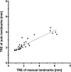
Results: TRE of AVG-AVG and phase-phase DIRs averaged 3.2 ± 1.0 and 2.6 ± 0.8 mm respectively (p < 0.001). Using AVG-AVG DIR, TREs for landmarks with <10 mm motion averaged 2.9 ± 2.0 mm, compared to 3.1 ± 1.9 mm for the remaining landmarks (p < 0.01). Comparatively, no significant difference was demonstrated for phase-phase DIRs. Dosimetrically, no significant difference in global dose metrics was observed between doses mapped with AVG-AVG DIR and the phase-phase DIR, but a positive linear relationship existed (p = 0.04) between the TRE of AVG-AVG DIR and local dose difference.
Conclusions: When the region of interest experiences <10 mm respiratory-induced motion, AVG-AVG DIR may provide sufficient geometric accuracy; conversely, extra attention is warranted, and phase-phase DIR is recommended. Dosimetrically, the differences in geometric accuracy between AVG-AVG and phase-phase DIRs did not impact global lung-based metrics. However, as more localized dose metrics are needed for toxicity assessment, phase-phase DIR may be required as its lower mean TRE improved voxel-based dosimetry.
Keywords: 4DCT; adaptive radiotherapy; deformable image registration accuracy; non-small cell lung cancer.
© 2021 The Authors. Journal of Applied Clinical Medical Physics published by Wiley Periodicals LLC on behalf of American Association of Physicists in Medicine.
Conflict of interest statement
Yulun He has nothing to disclose; Dr. Cazoulat has nothing to disclose; Dr. Wu has nothing to disclose; Dr. Peterson has nothing to disclose; Dr. McCulloch has nothing to disclose; Brian Anderson has nothing to disclose; Dr. Pollard‐Larkin has nothing to disclose; Dr. Balter reports grants from RaySearch Laboratories, grants from Varian Associates, outside the submitted work; Dr. Liao has nothing to disclose; Dr. Mohan has nothing to disclose; Dr. Brock reports grants from RaySearch Laboratories, Helen Black Image Guided Cancer Therapy Research Fund and Tumor Measurement Initiative of University of Texas MD Anderson Cancer Center, during the conduct of the study. In addition, Dr. Brock has a licensing agreement with RaySearch Laboratories with royalties paid.
Figures


References
-
- Hill RP, Bristow RG. Tumor and normal tissue response to radiotherapy. In: Tannock IF, Hill RP, Bristow RG, Harrington L, eds. The basic science of oncology, 5th. edn. New York: McGraw‐Hill Education Medical; 2016.
MeSH terms
Grants and funding
LinkOut - more resources
Full Text Sources
Medical

