

Evaluation of bone grafting for treatment of low-grade chondrosarcoma of long bones
- PMID: 34311593
- PMCID: PMC8320587
- DOI: 10.1177/03000605211025403
Evaluation of bone grafting for treatment of low-grade chondrosarcoma of long bones
Abstract
Objective: To retrospectively analyze the biological compatibility and oncologic outcomes of autogenous, allogeneic, or combined bone grafting.
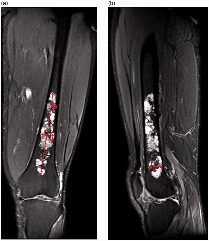
Methods: From April 2000 to December 2016, 37 patients with histologically confirmed low-grade intramedullary chondrosarcoma of the long bones at Kyungpook National University Hospital were enrolled in this retrospective study. All 37 patients underwent intralesional curettage (with or without cryotherapy) followed by bone grafting. Among the 24 patients who underwent cryotherapy, 13 were treated by prophylactic internal fixation (10 in the femur, 1 in the tibia, and 2 in the humerus). Thirteen patients underwent the same treatment without cryotherapy, whereas 12 did not undergo preventive internal fixation.
Results: A single intraoperative fracture was managed by plate fixation. One patient who underwent cryotherapy and internal fixation developed a fracture distal to the operation site 25 days after surgery, and this fracture was repaired with a long plate. None of the 37 patients showed any recurrence or metastasis.
Conclusions: Adequate intralesional curettage (with or without cryosurgery) combined with bone grafting using autogenous and allogeneic bone chips was effective for the treatment of low-grade intramedullary chondrosarcoma. Therefore, prophylactic internal fixation using a plate is recommended in the cryotherapy of definite cortical invasion in weight-bearing bones.
Keywords: Low-grade chondrosarcoma; bone graft; cryosurgery; curettage; internal fixation; long bone.
Conflict of interest statement
Figures


References
-
- Balasubramaniam S, Pawar V. Cytologic diagnosis of chondrosarcoma on fine needle aspiration cytology: a challenge! Journal of Krishna Institute of Medical Sciences University 2019; 8: 101–104. https://www.jkimsu.com/jkimsu-vol8no4/JKIMSU,%20Vol.%208,%20No.%204,%20O...
-
- Tsuda Y, Tsoi K, Stevenson JD, et al.. Development and external validation of nomograms to predict sarcoma-specific death and disease progression after surgical resection of localized high-grade conventional primary central chondrosarcoma and dedifferentiated chondrosarcoma. Bone Joint J 2020; 102-b: 1752–1759. - PubMed
-
- Blumstein G, Kelley B, Nelson S, et al.. Low-grade spinal malignancies: chordoma and chondrosarcoma. In: Singh K, Colman M. (eds) Surgical spinal oncology. Cham: Springer, 2020, pp.89–113. 10.1007/978-3-030-50722-0_7 - DOI
MeSH terms
LinkOut - more resources
Full Text Sources
Medical
Miscellaneous
