. 2021 Jul 26;11(1):15156.
doi: 10.1038/s41598-021-94314-1.

Extracellular NLRP3 inflammasome particles are internalized by human coronary artery smooth muscle cells and induce pro-atherogenic effects

Affiliations

Extracellular NLRP3 inflammasome particles are internalized by human coronary artery smooth muscle cells and induce pro-atherogenic effects

Susanne Gaul et al. Sci Rep. .

Abstract

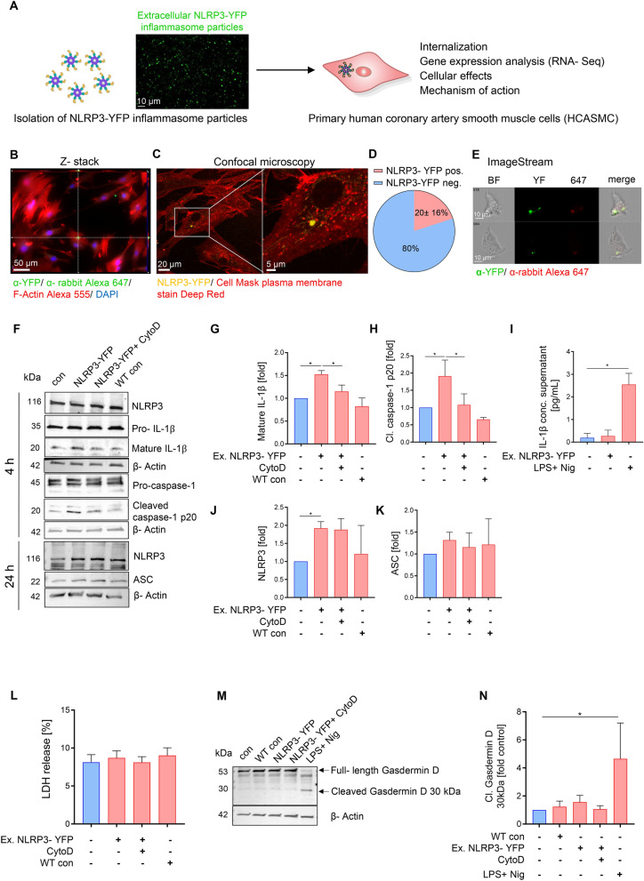
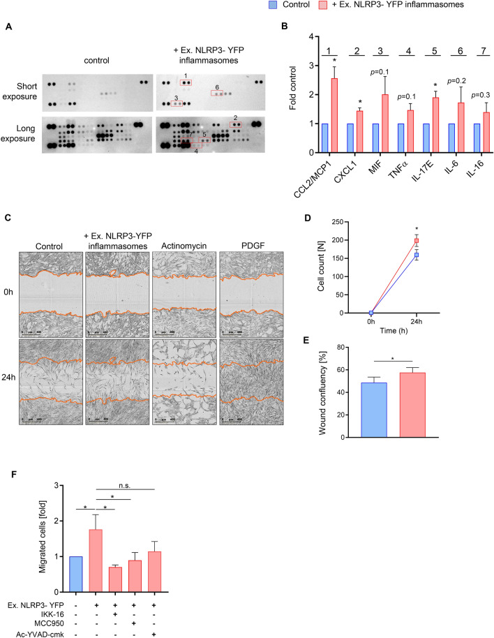
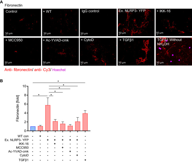
Inflammation driven by intracellular activation of the NLRP3 inflammasome is involved in the pathogenesis of a variety of diseases including vascular pathologies. Inflammasome specks are released into the extracellular compartment from disrupting pyroptotic cells. The potential uptake and function of extracellular NLRP3 inflammasomes in human coronary artery smooth muscle cells (HCASMC) are unknown. Fluorescently labeled NLRP3 inflammasome particles were isolated from a mutant NLRP3-YFP cell line and used to treat primary HCASMC for 4 and 24 h. Fluorescent and expressional analyses showed that extracellular NLRP3-YFP particles are internalized into HCASMC, where they remain active and stimulate intracellular caspase-1 (1.9-fold) and IL-1β (1.5-fold) activation without inducing pyroptotic cell death. Transcriptomic analysis revealed increased expression level of pro-inflammatory adhesion molecules (ICAM1, CADM1), NLRP3 and genes involved in cytoskleleton organization. The NLRP3-YFP particle-induced gene expression was not dependent on NLRP3 and caspase-1 activation. Instead, the effects were partly abrogated by blocking NFκB activation. Genes, upregulated by extracellular NLRP3 were validated in human carotid artery atheromatous plaques. Extracellular NLRP3-YFP inflammasome particles promoted the secretion of pro-atherogenic and inflammatory cytokines such as CCL2/MCP1, CXCL1 and IL-17E, and increased HCASMC migration (1.8-fold) and extracellular matrix production, such as fibronectin (5.8-fold) which was dependent on NFκB and NLRP3 activation. Extracellular NLRP3 inflammasome particles are internalized into human coronary artery smooth muscle cells where they induce pro-inflammatory and pro-atherogenic effects representing a novel mechanism of cell-cell communication and perpetuation of inflammation in atherosclerosis. Therefore, extracellular NLRP3 inflammasomes may be useful to improve the diagnosis of inflammatory diseases and the development of novel anti-inflammatory therapeutic strategies.


Conflict of interest statement

The authors declare no competing interests.

Figures

Figure 1
Figure 1
Extracellular NLRP3-YFP inflammasome particles are internalized by primary human coronary artery smooth muscle cells (HCASMC) and induce caspase-1 and IL-1β activation. (A) Schematic overview of study objective. Constitutively active mutant HEK NLRP3-YFP (p.D303N) cells were used for isolation of cell-free NLRP3-YFP inflammasome particles that are used for treatment of HCASMC (scale bar: 10 µm). (B) Internalization of extracellular NLRP3-YFP inflammasomes in HCASMC after 4 h of incubation determined by immunofluorescent staining (N = 3) with an anti-YFP antibody or IgG isotype control and fluorescently-labeled anti-rabbit Alexa 647 secondary antibody (scale bar: 50 µm). Alexa Fluor 555 Phalloidin and DAPI were used for F-Actin and nucleus staining, respectively. Z-stacks with xz and yz focal planes (white dashed lines) showing internalized NLRP3-YFP inflammasome in HCASMC. (C) Internalization of extracellular NLRP3-YFP inflammasomes (purified particles) (yellow). HCASMC were incubated for 4 h. Uptake was confirmed via confocal microscopy. Plasma membrane staining (red) was carried out using Cell Mask Deep Red (scale bar: 20 µm). Micrographs depicting internalization and subcellular localisation of NLRP3-YFP inflammasome particles are shown in overview and enhanced magnification (scale bar: 5 µm). (D) For quantification, four representative fields of view were analyzed. (E) Representative ImageStream analysis of HCASMC showing internalized extracellular NLRP3-YFP inflammasome (scale bar: 10 µm) by merging the brightfield image (BF), YFP signal of the NLRP3-YFP inflammasome and the immunostained signal of the anti-YFP/ anti-rabbit Alexa 647 antibody. (F-K) HCASMC were treated with extracellular NLRP3-YFP inflammasome particles (3:1) for 4 h and 24 h with our without pre-incubation of Cytochalasin D (CytoD, 4 µM) for 30 min and total protein lysate was used for immunoblotting. Immunoblot (N = 4) of (F) NLRP3, pro-IL-1β and mature IL-1β 17 kDa, pro-caspase-1 and activated caspase-1 p20 (4 h) as well as endogenous NLRP3 and ASC after 24 h. The corresponding densitometric analysis of (G) mature cleaved IL-1β and (H) activated caspase-1 p20 as well as (I) IL-1β secretion (pg/ml) is shown (after 4 h). As positive control for endogenous inflammasome activation and IL-1β secretion, cells were stimulated with LPS (1 µg/ml, 3 h) and Nigericin (10 µM, 90 min). (J, K) Densitometric analysis of NLRP3 and ASC protein level after 24 h of stimulation with extracellular NLRP3-YFP inflammasome particles (N = 4). β-Actin was used as housekeeping control. To exclude effects of particle isolation, same protocol for inflammasome particle isolation was performed on WT HEK cells and were used for treatment (WT con) of HCASMC. Data were normalized on β-Actin and set at 1. (L) Membrane disruption was measured by the release of lactate dehydrogenase (LDH) into supernatant of HCASMC treated with NLRP3-YFP inflammasome particles (3:1 particles/cell) for 4 h. LDH values were normalized to positive LDH control which was set 100%. Immunoblot (N = 3) of (M) Gasdermin D (full-length 53 kDa) and its active cleaved N-terminal fragment (30 kDa). β-Actin was used as housekeeping control. (N) Densitometric analysis of the cleaved Gasdermin D fragment (30 kDa) was performed and normalized on untreated control which was set at 1. HCASMC treated with LPS + Nig were used as positive control. Differences between the groups were analysed by One-way ANOVA and uncorrected Fishers LSD post-hoc test (*p < 0.05).
Figure 2
Figure 2
Extracellular NLRP3-YFP inflammasome particles regulate gene expression in HCASMC. (A) Heatmap of genes upregulated in HCASMC treated with extracellular NLRP3-YFP inflammasome particles (N = 2) for 24 h. (B) List of the ten most upregulated genes. (C) Validation of four listed genes (NLRP3, ICAM1, CADM1, SPON1) using qPCR after 4 and 24 h treatment with extracellular NLRP3-YFP particles. Data were normalized on the mean of two housekeeping genes (RPLP0 and TBP) and were log2-transformed for equal distribution. Untreated cells (control) were set at 1. (D) Gene enrichment analysis of the 200 most upregulated genes (mean log2 fold > 3.5 vs. control) was performed using EnrichR and NCATS BioPlanet 2019 comprehensive integrated pathway analysis. Pathways with p < 0.05 are shown. mRNA expression of (E) adhesion molecule ICAM1 and (F) NLRP3 in HCASMC (N = 9) treated with extracellular NLRP3-YFP inflammasomes (3:1 particles/ cell) for 24 h with or without pre-incubation of caspase-1 inhibitor (Ac-YVAD-cmk, 25 µg/ml), NLRP3 inhibitor (MCC950, 1 µM) and NFκB inhibitor (IKK-16, 2 µM). Data are expressed as mean ± SEM and were normalized on the mean of two housekeeping genes (RPLP0 and TBP) and referenced to untreated control which was set at 1. Differences between the groups were calculated using One-way ANOVA and uncorrected Fisher´s LSD post-hoc test (*p < 0.05). (G) Selected genes (NLRP3, ICAM1, CADM1, NUP210) were re-analysed using a pre-existing microarray dataset and the gene expression was compared between atheroma plaque and adjacent macroscopically intact tissue from the same patient (N = 34). Data are expressed as mean ± SEM and differences between the groups were calculated using unpaired, two-tailed Student´s T-Test (***p < 0.001, ****p < 0.0001).
Figure 3
Figure 3
Extracellular NLRP3-YFP inflammasome particles promote release of pro-atherogenic cytokines and cell migration in HCASMC. (A, B) Supernatant of HCASMC (N = 3) after 4 h treatment with extracellular NLRP3-YFP inflammasomes (3:1 particles/cell) were used for the human proteome profiler cytokine array. Modified cytokines/chemokines were surrounded with a red square and labelled (1–7). Membranes were applied for short and long-time exposure. (B) Densitometric analysis of the labelled cytokines (1–7) after background subtraction was performed. Data were normalized on supernatant from control cells which was set at 1. (C) Scratch assay of HCASMC treated with NLRP3-YFP inflammasome particles (3:1 particles/ cell) for 4 h. PDGF (10 ng/ml) and Actinomycin (5 µg/ml) were stimulated for 24 h. Representative images of wounds at the beginning (0 h) and after 24 h are shown. Wounds were automatically acquired within the CO2 incubator using the IncuCyte ZOOM software package. (D) Cell count [N] at 0 h and after 24 h within the scratch and (E) wound confluency [%] was calculated using the IncuCyte ZOOM software. Data are expressed as mean ± SEM and differences between the groups were calculated using unpaired, two-tailed Student´s T-Test (*p < 0.05). (F) Smooth muscle cell migration was studied in a Boyden chamber by adding NLRP3-YFP inflammasome particles (3:1 particles/ cell) to HCASMC for 4 h and analyzing the migrated cells on the lower side of the insert membrane which was stained with crystal violet and measured at 560 nm in a fluorometer (N = 4–6). Differences between the groups were analysed by One-way ANOVA and uncorrected Fishers LSD post-hoc test (*p < 0.05).
Figure 4
Figure 4
Extracellular NLRP3-YFP inflammasome particles induce extracellular matrix (ECM) production in HCASMC that can be reduced by different inhibitors. (A) Immunofluorescent staining of cell-free ECM protein fibronectin (red) produced by HCASMC after treatment with extracellular NLRP3-YFP inflammasomes (3:1 particles/ cell) for 24 h (N = 5). ECM production was reduced when cells were co-stimulated with inhibitors such as IKK-16 (2 µM), Ac-YVAD-cmk (25 µg/ml), MCC950 (1 µM) for 24 h or pre-incubation with CytoD (2.5 µg/ml) for 30 min. Antibody against fibronectin (1 µg/ml, Santa Cruz) or IgG mouse isotype control as well as anti-rabbit anti-mouse Cy3 antibody (1:1000, Dianova) were used (scale bar: 50 µm). Ammonium hydroxide (NH4OH) was used to remove cells and only stain extracellular fibronectin. Negative Hoechst (nucleus) staining showed successful removal of cells. TGFβ1 (48 h) was used as positive control. (B) Five random selected fields were used for ImageJ analysis and the positive stained area (% of area) from images with same magnification was calculated. Data were normalized on the untreated control which was set at 1. Data are expressed as mean ± SEM and differences between the groups were compared using One-way ANOVA and Sidak´s post-hoc test (*p < 0.05).

References

    1. Chen GY, Nuñez G. Sterile inflammation: sensing and reacting to damage. Nat. Rev. Immunol. 2010;10:826–837. doi: 10.1038/nri2873. - DOI - PMC - PubMed
    1. Zewinger S, et al. Apolipoprotein C3 induces inflammation and organ damage by alternative inflammasome activation. Nat. Immunol. 2020;21:30–41. doi: 10.1038/s41590-019-0548-1. - DOI - PubMed
    1. Schroder K, Tschopp J. The inflammasomes. Cell. 2010;140:821–832. doi: 10.1016/j.cell.2010.01.040. - DOI - PubMed
    1. Broz P, Pelegrín P, Shao F. The gasdermins, a protein family executing cell death and inflammation. Nat. Rev. Immunol. 2020;20:143–157. doi: 10.1038/s41577-019-0228-2. - DOI - PubMed
    1. Yin, Y. et al. Early hyperlipidemia promotes endothelial activation via a caspase-1-sirtuin 1 pathway. Arteriosclerosis, Thrombosis Vasc. Biol.35, 804–816 (2015). - PMC - PubMed

Publication types

MeSH terms