. 2021 Oct;58(10):5356-5368.
doi: 10.1007/s12035-021-02485-9. Epub 2021 Jul 27.

Genetic Exchange of Lung-Derived Exosome to Brain Causing Neuronal Changes on COVID-19 Infection

Affiliations

Genetic Exchange of Lung-Derived Exosome to Brain Causing Neuronal Changes on COVID-19 Infection

Shiek S S J Ahmed et al. Mol Neurobiol. 2021 Oct.

Abstract

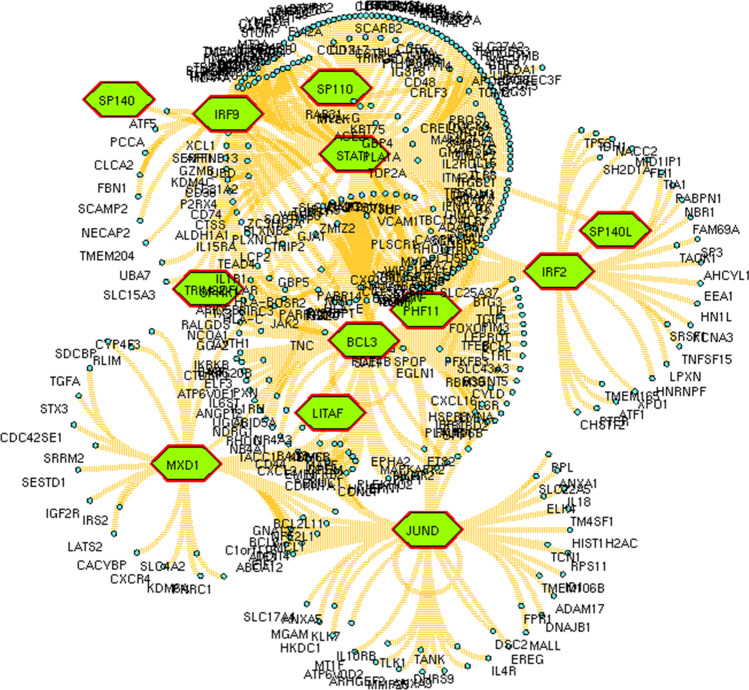
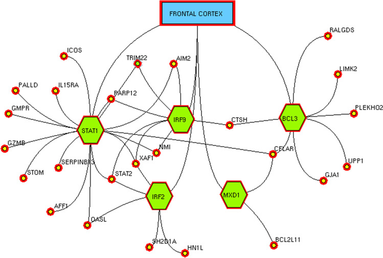
The pandemic of novel coronavirus 2 (SARS-CoV-2) has made global chaos for normal human living. Despite common COVID-19 symptoms, variability in clinical phenotypes was reported worldwide. Reports on SARS-CoV-2 suggest causing neurological manifestation. In addition, the susceptibility of SARS-CoV-2 in patients with neurodegenerative diseases and its complexity are largely unclear. Here, we aimed to demonstrate the possible transport of exosome from SARS-CoV-2-infected lungs to the brain regions associated with neurodegenerative diseases using multiple transcriptome datasets of SARS-CoV-2-infected lungs, RNA profiles from lung exosome, and gene expression profiles of the human brain. Upon transport, the transcription factors localized in the exosome regulate genes at lateral substantia nigra, medial substantia nigra, and superior frontal gyrus regions of Parkinson's disease (PD) and frontal cortex, hippocampus, and temporal cortex of Alzheimer's disease (AD). On SARS-CoV-2 infection, BCL3, JUND, MXD1, IRF2, IRF9, and STAT1 transcription factors in the exosomes influence the neuronal gene regulatory network and accelerate neurodegeneration. STAT1 transcription factor regulates 64 PD genes at lateral substantia nigra, 65 at superior frontal gyrus, and 19 at medial substantia nigra. Similarly, in AD, STAT1 regulates 74 AD genes at the temporal cortex, 40 genes at the hippocampus, and 16 genes at the frontal cortex. We further demonstrate that dysregulated neuronal genes showed involvement in immune response, signal transduction, apoptosis, and stress response process. In conclusion, SARS-CoV-2 may dysregulate neuronal gene regulatory network through exosomes that attenuate disease severity of neurodegeneration.

Keywords: Alzheimer’s disease; Covid-19; Exosome; Neurodegeneration; Parkinson’s disease; SARS‐CoV‐2.


Conflict of interest statement

The authors declare no competing interests.

Figures

Fig. 1
Fig. 1
Exosomal communication network. Network representing the exosomal connectivity between lungs to the neuronal regions associated with neurodegenerative diseases through the blood–brain barrier (BBB). The lungs are represented as violet node defining connection with the BBB (green node) by bold dotted edges. BBB connects various brain regions associated with PD (yellow) and AD (red). The gray-colored nodes represent the neurons, orange nodes represent astrocyte, and blue nodes represent microglia cells
Fig. 2
Fig. 2
Co-expressed network of neurodegenerative disease genes with TFs. Network representing the co-expressed up-regulated genes (blue ellipse) in PD and AD along with the 19 transcription factors (green polygon) transported from the lung through exosome on COVID-19 infection
Fig. 3
Fig. 3
Regulatory network in lateral substantia nigra. Network representing the transcription factors (green polygon) regulating (back dotted edges) the up-regulated genes (yellow ellipse) in Parkinson’s disease
Fig. 4
Fig. 4
Regulatory network in medial substantia nigra. Network representing the transcription factors (green polygon) regulating (back dotted edges) the up-regulated genes (yellow ellipse) in Parkinson’s disease
Fig. 5
Fig. 5
Regulatory network in the frontal gyrus. Network representing the transcription factors (green polygon) regulating (back dotted edges) the up-regulated genes (yellow ellipse) in Parkinson’s disease
Fig. 6
Fig. 6
Regulatory network in the frontal cortex. Network representing the transcription factors (green polygon) regulating (back dotted edges) the up-regulated genes (yellow ellipse) in Alzheimer’s disease
Fig. 7
Fig. 7
Regulatory network in the hippocampus. Network representing the transcription factors (green polygon) regulating (back dotted edges) the up-regulated genes (yellow ellipse) in Alzheimer’s disease
Fig. 8
Fig. 8
Regulatory network in the temporal cortex. Network representing the transcription factors (green polygon) regulating (back dotted edges) the up-regulated genes (yellow ellipse) in Alzheimer’s disease
Fig. 9
Fig. 9
Functional enrichment of Parkinson’s disease genes regulated by the transcription factors. Transcription factors activating PD genes showing major involvement in the immune system, signal transduction, cell cycle, and programmed cell death
Fig. 10
Fig. 10
Functional enrichment of Alzheimer’s disease genes regulated by the transcription factors. Transcription factors activating AD genes showing significant involvement in the immune system, signal transduction, metabolism, cell cycle, and response to external stimuli

References

    1. Colombo M, Raposo G, Théry C. Biogenesis, secretion, and intercellular interactions of exosomes and other extracellular vesicles. Annu Rev Cell Dev Biol. 2014;30:255–289. doi: 10.1146/annurev-cellbio-101512-122326. - DOI - PubMed
    1. Statello L, Maugeri M, Garre E, Nawaz M, Wahlgren J, Papadimitriou A, Lundqvist C, Lindfors L, et al. Identification of RNA-binding proteins in exosomes capable of interacting with different types of RNA: RBP-facilitated transport of RNAs into exosomes. PLoS One. 2018;13(4):e0195969. doi: 10.1371/journal.pone.0195969. - DOI - PMC - PubMed
    1. Johnstone RM, Adam M, Hammond JR, Orr L, Turbide C. Vesicle formation during reticulocyte maturation. Association of plasma membrane activities with released vesicles (exosomes) J Biol Chem. 1987;262(19):9412–20. doi: 10.1016/S0021-9258(18)48095-7. - DOI - PubMed
    1. Ludwig N, Whiteside TL, Reichert TE. Challenges in exosome isolation and analysis in health and disease. Int J Mol Sci. 2019;20(19):4684. doi: 10.3390/ijms20194684. - DOI - PMC - PubMed
    1. Zhao W, Zheng XL, Zhao SP. Exosome and its roles in cardiovascular diseases. Heart Fail Rev. 2015;20(3):337–348. doi: 10.1007/s10741-014-9469-0. - DOI - PubMed