Review
. 2021 Nov;22(1_suppl):71-83.
doi: 10.1177/11297298211018062. Epub 2021 Jul 27.

Ultrasound evaluation of access complications: Thrombosis, aneurysms, pseudoaneurysms and infections

Affiliations
Review

Ultrasound evaluation of access complications: Thrombosis, aneurysms, pseudoaneurysms and infections

Mario Meola et al. J Vasc Access. 2021 Nov.

Abstract

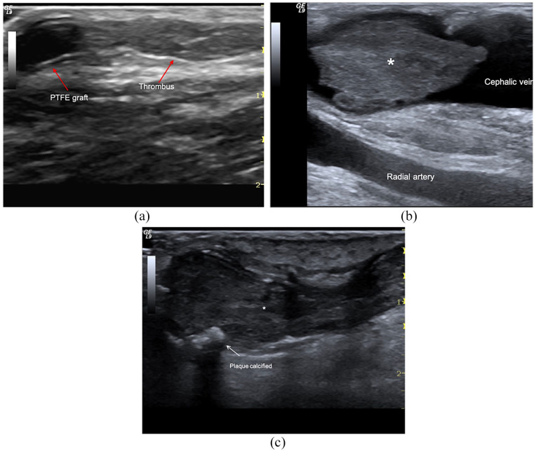
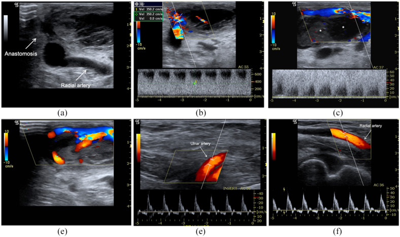
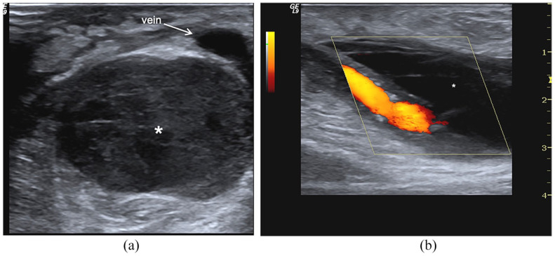
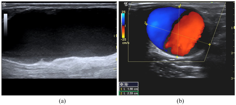
Arteriovenous fistula (AVF) complications are classified based on fistula outcomes. This review aims to update colour Doppler (CD) and pulse wave Doppler (PWD) roles in managing early and late complications of the native and prosthetic AVF. Vascular access (VA) failure occurs because inflow or outflow stenosis activates Wirchow's triad inducing thrombosis. Therefore, the diagnosis of the tributary artery and outgoing vein stenosis will be the first topic considered. Post-implantation complications occur from the inability to achieve AVF maturation and dialysis suitability due to inflow/outflow stenosis. Late stenosis is usually a sequence of early defects repaired to maintain patency. Less frequently, in the mature AVF or graft, complications are acquired 'de novo'. They derive either from incorrect management of vascular access (haematoma, pseudoaneurysm, prosthesis infection) or wall pathologies (aneurysm, myxoid valve degeneration, kinking, coiling, abnormal dilation from defects of elastic structures). High-resolution transducers (10-20 MHz) allow the characterization of the wall damage, haemodynamic dysfunctions, early and late complications even if phlebography remains the gold standard for the diagnosis for its sensitivity and specificity.

Keywords: AVF complications; doppler imaging; fistula aneurysm; inflow/outflow stenosis; pseudoaneurysm; vascular access thrombosis.


Conflict of interest statement

Declaration of conflicting interests: The author(s) declared no potential conflicts of interest with respect to the research, authorship, and/or publication of this article.

Figures

Figure 1.
Figure 1.
Geometric stenosis. B-Mode evaluates reduction of the vessel lumen for a atheromatous plaque either as a percentage reduction in the linear diameter (A/B × 100 = % stenosis) (a, b), or as a percentage reduction in the cross-sectional area ((A2/B2) × 100 = % stenosis)) calculated from the diameter values (c) or cross-sectional areas (d). Angiography defines ‘critical’ as a reduction in diameter >50%.
Figure 2.
Figure 2.
Outflow stenosis. (a) B-Mode image of an iuxta-anastomotic tract of a mature distal radio-cephalic fistula. Stenosis is 2.12 mm in length and derives from reactive neointimal hyperplasia is represented in the image by the mid-level parietal echoes, (b) PWD sampling of the same fistula. Aliasing and bruit colour are evident at CD, while the spectral curve shows PSV> 350 cm/s, DV >180 cm/s, spectral broadening and aliasing, (c) Power Doppler sampling of a brachio-cephalic AVG. The graft flow rate is reduced (450 mL/min) because stenosis is located at the venous anastomosis level (PSV 370 cm/s, DV 240 cm/s) (d).
Figure 3.
Figure 3.
Inflow stenosis. (a) anastomotic breach CD sampling of a 1-month native side-to-end radio-cephalic fistula, (b) brachial flow rate calculation = 284 mL/min, and (c) spectral analysis at the level of breach anastomosis reveals severe stenosis: PSV > 400 cm/s, EDV 280 cm/s, spectral broadening, wall-thump, bruit colour and aliasing (Doppler angle = 28°).
Figure 4.
Figure 4.
Tributary artery stenosis. Male, 78 years old with maturation defect of distal radio-cephalic fistula 24 days after implantation: (a) Spectral Doppler highlights segmental stenosis of the tributary artery. Colour Doppler sampling shows aliasing and colour bruit; spectral analysis shows an explosive increase of PSV (>300 cm/s), and EDV at the level of stenosis (Doppler angle 36°), spectral broadening and aliasing; (b) downstream PSV is 130 cm/s and the peak-velocity ratio is 2 compared to the stenosis. (c) Upstream, the peak-velocity ratio is >2.5, but the spectral amplitude is elevated. Note that the Doppler angle is at 36°, so not perfectly aligned with the vessel’s longitudinal axis; this technical mistake underestimated the PSV.
Figure 5.
Figure 5.
AVF thrombosis: (a) Brachio-cephalic graft. Occlusive clot (*) in the cephalic vein, (b) distal radio-cephalic fistula. Acute non-occlusive thrombosis of anastomosis and outgoing vein, and (c) acute obstructive thrombosis (<24 h) (*) of cephalic vein in mature AVF. Wall calcifications (arrows) are also evident.
Figure 6.
Figure 6.
Early failure of radiocephalic AVF for inflow stenosis and thrombosis. AVF malfunction appears in the first HD session. (a) B-Mode, CD and PWD sampling, (b) highlight a sub-occlusive thrombosis (*) of the venous effluent favoured by severe inflow stenosis (aliasing, PSV > 600 cm/s, Doppler angle 55°). There is a thin jet between the thrombus and vein. The spectral trace of radial, (d) and ulnar artery reveals a high resistance velocity/time curve due to complete obstruction of anastomosis. The patient carried out percutaneous declotting even if the stenosis was not critical because the brachial artery flow rate was still >650 mL/min. (d) Distal mature AVF. Acute and obstructive thrombosis (<24 h) (*) of the cephalic vein. At B-Mode, there are also wall calcifications (arrows). Spectral trace of ulnar (e) radial (f) arteries reveals a high resistance velocity/time curve due to complete obstruction of anastomosis.
Figure 7.
Figure 7.
Haematoma: (a) At B-Mode, perivascular haematoma appears as a complex hypoechoic and corpuscolated fluid collection with debris and fibrin striae (*), (b) at CD sampling, it is not perfused (*) and surrounds the draining vein.
Figure 8.
Figure 8.
Aneurysm: (a) B-Mode long-axis scans of the cephalic vein. The true aneurysm appears as a fusiform or saccular vein dilation, (b) short axis of the vein. A large red-blue vortex similar to the ‘Korean flag’ is evident at CD/PD sampling in the aneurysmal sac.
Figure 9.
Figure 9.
Arterial pseudoaneurysm: (a) B-Mode scans. A circular swirling in a brachial artery pseudoaneurysm is evident. The spontaneous blood echogenicity is due to the slowdown of blood in the pseudoaneurysm. At CD, the recirculation of blood draws the ‘yin-yang’ sign, (b) spectral Doppler in correspondence of the collar, shows a ‘to and fro’ pattern (isodiphasic deflection).
Figure 10.
Figure 10.
Venous pseudoaneurysm. A fresh and not very large pseudo-aneurysm can be treated with compression of the breach using the transducer. Ultrasound-guided percutaneous injection of bovine thrombin (100–1500 IU) promotes a rapid thrombosis of the pseudo-aneurysm.
Figure 11.
Figure 11.
Calcifications: (a) Well-functioning long-term AVF with coarse calcifications of the anastomotic chamber, (b) chronic calcified thrombus in a non-functioning fistula.

References

    1. Gallieni M, Hollenbeck M, Inston N, et al.. Clinical practice guideline on peri- and postoperative care of arteriovenous fistulas and grafts for haemodialysis in adults. Nephrol Dial Transplant 2019; 34(Supplement 2): ii1–ii42. - PubMed
    1. Schmidli J, Widmer MK, Basile C, et al.. Editor’s choice - vascular access: 2018 clinical practice guidelines of the European Society for Vascular Surgery (ESVS). Eur J Vasc Endovasc Surg 2018; 55: 757–818. - PubMed
    1. Schinstock CA, Albright RC, Williams AW, et al.. Outcomes of arteriovenous fistula creation after the Fistula First Initiative. Clin J Am Soc Nephrol 2011; 6:1996–2002. - PMC - PubMed
    1. McGrogan D, Al Shakarchi J, Khawaja A, et al.. Arteriovenous fistula outcomes in the elderly. J Vasc Surg 2015; 62:1652–1657. - PubMed
    1. Roy-Chaudhury P, Spergel LM, Besarab A, et al.. Biology of arteriovenous fistula failure. J Nephrol 2007; 20:150–163. - PubMed