Multicenter Study
. 2022 Jan;77(1):282-295.
doi: 10.1111/all.15025. Epub 2021 Aug 10.

Adverse COVID-19 outcomes in immune deficiencies: Inequality exists between subclasses

Affiliations
Multicenter Study

Adverse COVID-19 outcomes in immune deficiencies: Inequality exists between subclasses

Elif Karakoc Aydiner et al. Allergy. 2022 Jan.

Abstract

Background: Genetic deficiencies of immune system, referred to as inborn errors of immunity (IEI), serve as a valuable model to study human immune responses. In a multicenter prospective cohort, we evaluated the outcome of SARS-CoV-2 infection among IEI subjects and analyzed genetic and immune characteristics that determine adverse COVID-19 outcomes.

Methods: We studied 34 IEI patients (19M/15F, 12 [min: 0.6-max: 43] years) from six centers. We diagnosed COVID-19 infection by finding a positive SARS-CoV-2 PCR test (n = 25) and/or a lung tomography scoring (CORADS) ≥4 (n = 9). We recorded clinical and laboratory findings prospectively, fitted survival curves, and calculated fatality rates for the entire group and each IEI subclass.

Results: Nineteen patients had combined immune deficiency (CID), six with predominantly antibody deficiency (PAD), six immune dysregulation (ID), two innate immune defects, and one in the autoinflammatory class. Overall, 23.5% of cases died, with disproportionate fatality rates among different IEI categories. PAD group had a relatively favorable outcome at any age, but CIDs and IDs were particularly vulnerable. At admission, presence of dyspnea was an independent risk for COVID-related death (OR: 2.630, 95% CI; 1.198-5.776, p < .001). Concerning predictive roles of laboratory markers at admission, deceased subjects compared to survived had significantly higher CRP, procalcitonin, Troponin-T, ferritin, and total-lung-score (p = .020, p = .003, p = .014, p = .013, p = .020; respectively), and lower absolute lymphocyte count, albumin, and trough IgG (p = .012, p = .022, p = .011; respectively).

Conclusion: Our data disclose a highly vulnerable IEI subgroup particularly disadvantaged for COVID-19 despite their youth. Future studies should address this vulnerability and consider giving priority to these subjects in SARS-Cov-2 therapy trials.

Keywords: COVID-19; SARS-Cov-2; inborn errors of immunity; outcome.


Conflict of interest statement

All authors declare that they have no conflict of interest related to this work.

Figures

FIGURE 1
FIGURE 1
Distribution of IEI categories and the probability of survival following COVID‐19. (A) A pie‐chart displays of the IEI categories and corresponding subclasses of the study group. AED‐ID: anhidrotic ectodermal dysplasia‐immune dysregulation, AGAM, agammaglobulinemia; AUTOINF, autoinflammatory; CID, combined immunodeficiency; CMC, chronic mucocutaneous candidiasis; CVID, common variable immunodeficiency; CORADS, COVID‐19 Reporting and Data System; COVID‐19, coronavirus infectious disease 2019; DNARD, DNA repair defects; EBV‐s, Epstein‐Barr virus susceptibility; F‐HLH, familial hemophagocytic lymphohistiocytosis; HIES, hyperimmunoglobulin E syndrome; HIGM, hyperimmunoglobulin M syndrome; HPV‐S, human papillomavirus susceptibility; ID, immune dysregulation; IID, innate immune defect; Type I IFN, Type I interferonopathy; NA, not available; PAD, predominantly antibody deficiency; PCR, polymerase chain reaction; PID, primary immune deficiency; SCID, severe combined immunodeficiency; SCID, syndromic combined immunodeficiency; Synd of autoimm, syndromes of autoimmunity. (B–D) Kaplan‐Meier curves showing the probability of survival as a function of days following hospital admission for the entire study group or the subgroups indicated in each graph
FIGURE 2
FIGURE 2
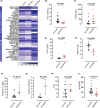
Clinical and laboratory characteristics of the study group concerning COVID‐19 outcome. (A) Heatmap display of the comparative characteristics of deceased vs. survived patients. The percentage of each parameter for the overall study population, or the indicated groups, is transformed into a color according to the scale indicated at the bottom. A Fisher's exact test compared the two groups for statistical significance. *p = .039, **p < .001, ***p = .17, #: number, BCG, Bacilli Calmette Guerin; CSB, class‐switched B cell; CD, clusters of differentiation; CORADS, COVID‐19 Reporting and Data System; IBD, inflammatory bowel disease; Ig, immunoglobulin; LDH, lactate dehydrogenase; LE, liver enzymes (SGOT and SGPT); NK, natural killer; PAD; primary antibody deficiency; RFT, renal function tests (creatinine and blood urea nitrogen); RTE, recent thymic emigrants; SARS‐CoV2 PCR, severe acute respiratory syndrome coronavirus 2 polymerase chain reaction, (B) Absolute lymphocyte count (ALC), (C) C reactive protein (CRP), (D) Procalcitonin (PcT), (E) Albumin, (F) Troponin‐T, (G) Ferritin, (H) Total lung score (TLS), and (I) Immunoglobulin G (IgG). In B through I, the error bars represent the median and IQR values, and the statistics used the Mann‐Whitney‐U test
FIGURE 3
FIGURE 3
Peripheral blood counts and immune subsets of deceased vs. survived subjects. (A) Absolute neutrophil count (ANC), (B) eosinophil, (C) monocyte counts concomitant with COVID infection, (D) T‐cell counts, (E) helper T‐cell counts, (F) B‐cell count, (G) natural killer (NK) cell count, (H) class‐switched memory B cell (CSB) %, and (I) recent thymic emigrant (RTE) %. Red symbols indicate deceased subjects, and the black symbols those survived COVID. The gray shaded area demarcated by dotted lines shows the age‐specific normal range
FIGURE 4
FIGURE 4
Longitudinal assessment of laboratory investigations of IEI patients who were succumbed to COVID. (A) Absolute lymphocyte count (ALC), (B) absolute neutrophil count (ANC), (C) eosinophil, (D) monocyte, (E) C reactive protein (CRP), (F) procalcitonin (PcT), (G) albumin, (H) troponin T and (I) ferritin levels. Horizontal gray bars indicate the upper and lower range. *pre‐COVID vs. per‐COVID albumin; p = .043, (Wilcoxon test)

References

    1. WHO Coronavirus disease (COVID‐19) pandemic. 2020; https://www.WHO.int/emergencies/diseases/novel‐coronavirus‐2019, 2020. Accessed September 03, 2020.
    1. Li Q, Guan X, Wu P, et al. Early transmission dynamics in Wuhan, China, of novel coronavirus‐infected pneumonia. N Engl J Med. 2020;382(13):1199‐1207. - PMC - PubMed
    1. Zhu N, Zhang D, Wang W, et al. A novel coronavirus from patients with pneumonia in China, 2019. N Engl J Med. 2020;382(8):727‐733. - PMC - PubMed
    1. Chen G, Wu D, Guo W, et al. Clinical and immunological features of severe and moderate coronavirus disease 2019. J Clin Invest. 2020;130(5):2620‐2629. - PMC - PubMed
    1. Levin AT, Hanage WP, Owusu‐Boaitey N, Cochran KB, Walsh SP, Meyerowitz‐Katz G. Assessing the age specificity of infection fatality rates for COVID‐19: systematic review, meta‐analysis, and public policy implications. Eur J Epidemiol. 2020;35(12):1123‐1138. - PMC - PubMed

Publication types