. 2021 Aug 19;184(17):4547-4563.e17.
doi: 10.1016/j.cell.2021.07.003. Epub 2021 Jul 26.

ELAVL4, splicing, and glutamatergic dysfunction precede neuron loss in MAPT mutation cerebral organoids

Affiliations

ELAVL4, splicing, and glutamatergic dysfunction precede neuron loss in MAPT mutation cerebral organoids

Kathryn R Bowles et al. Cell. .

Abstract

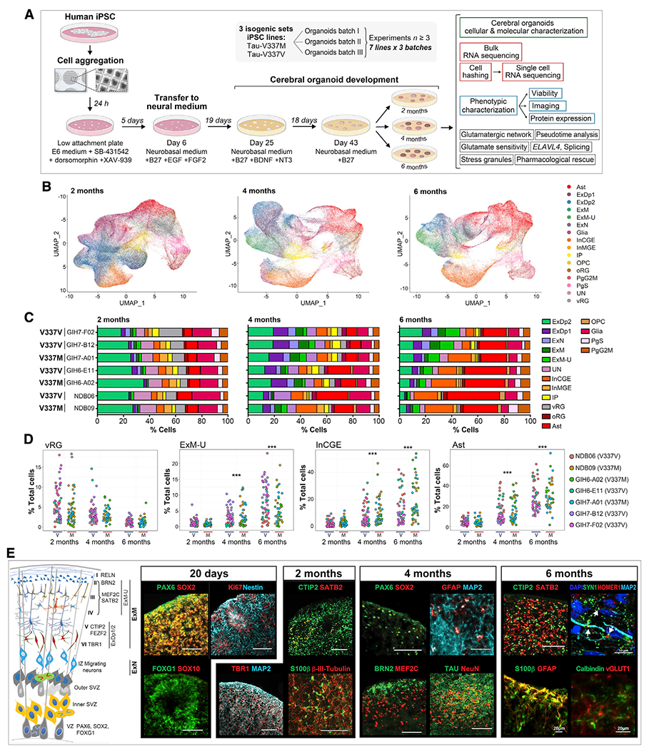
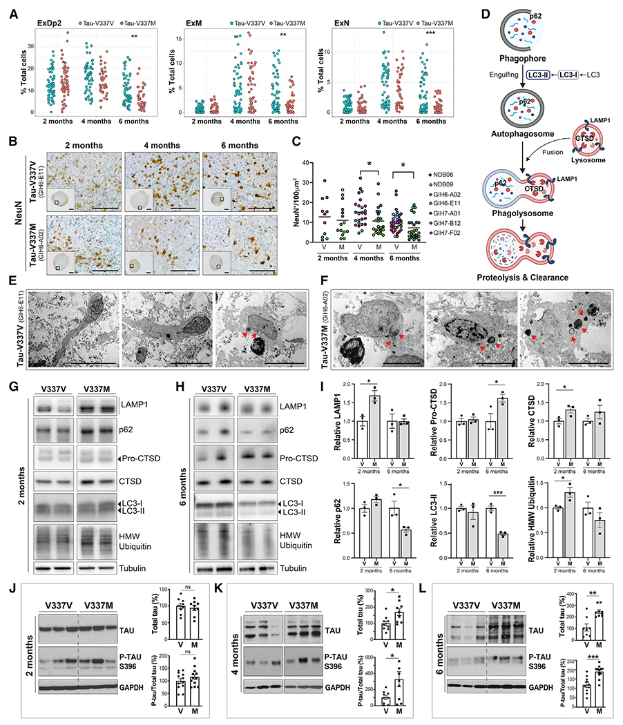
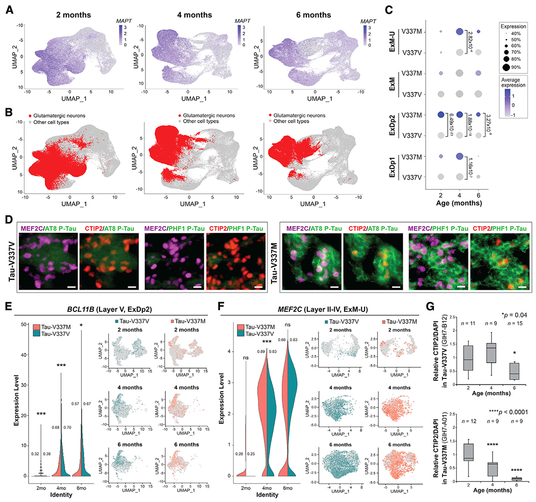
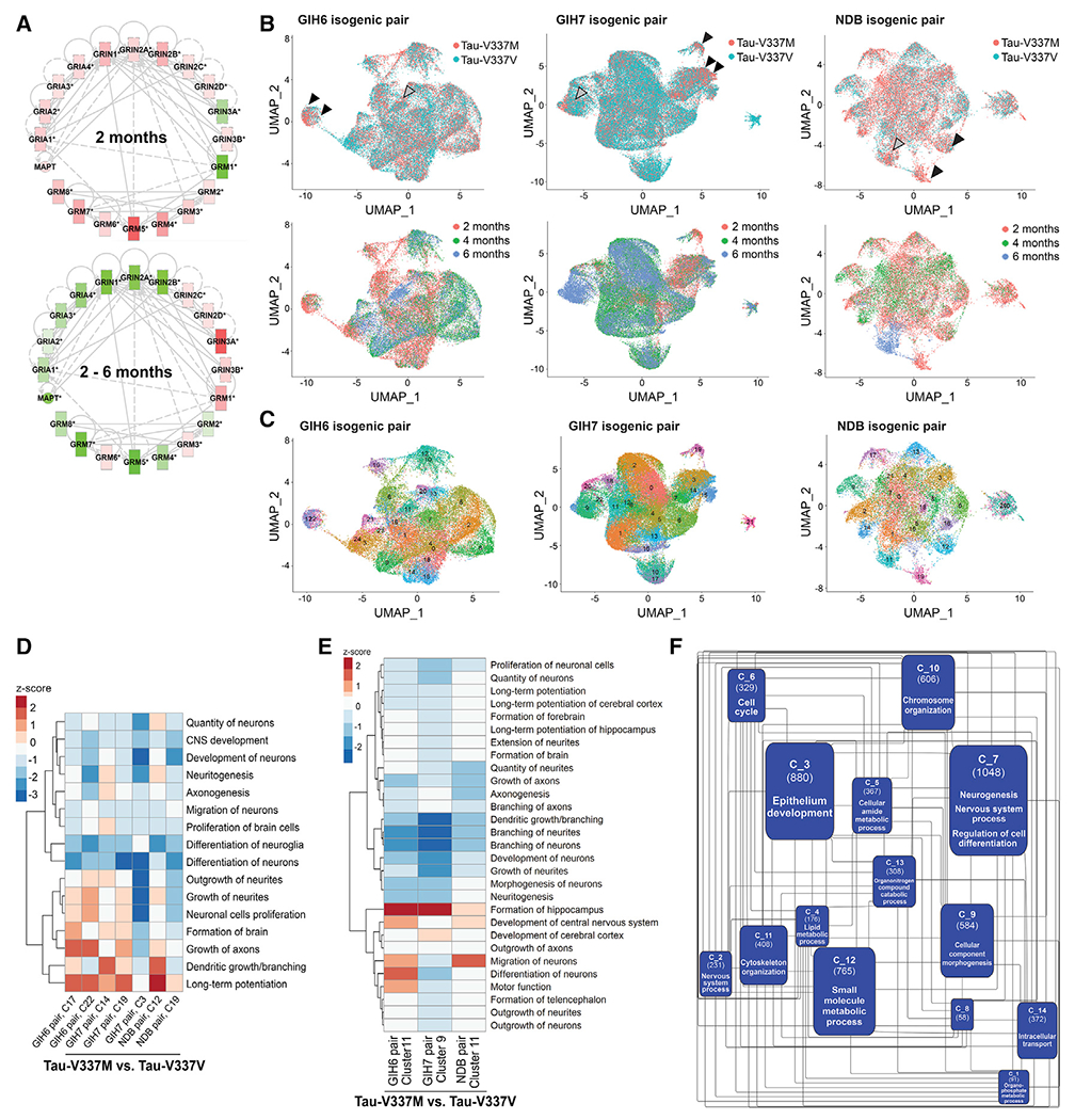
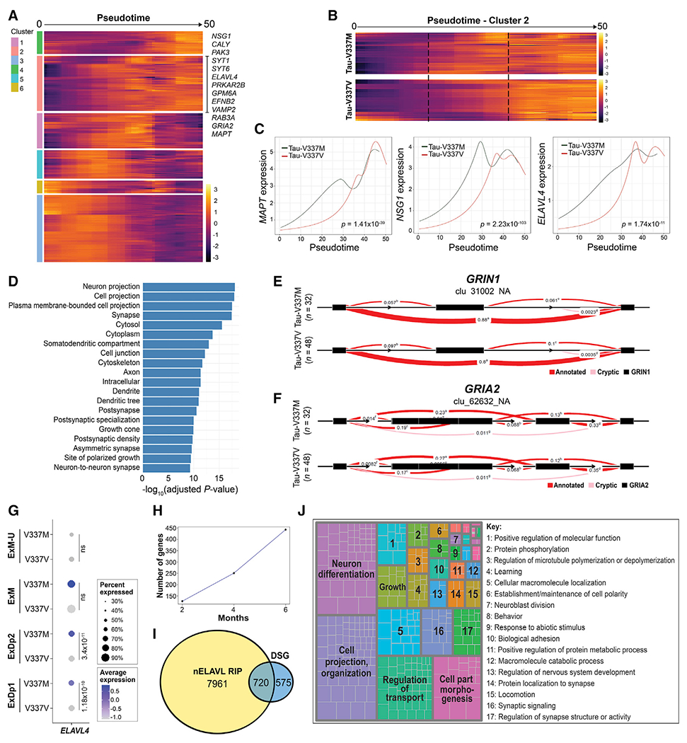
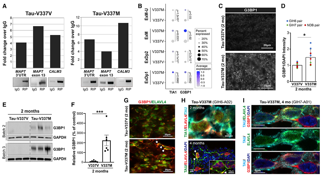
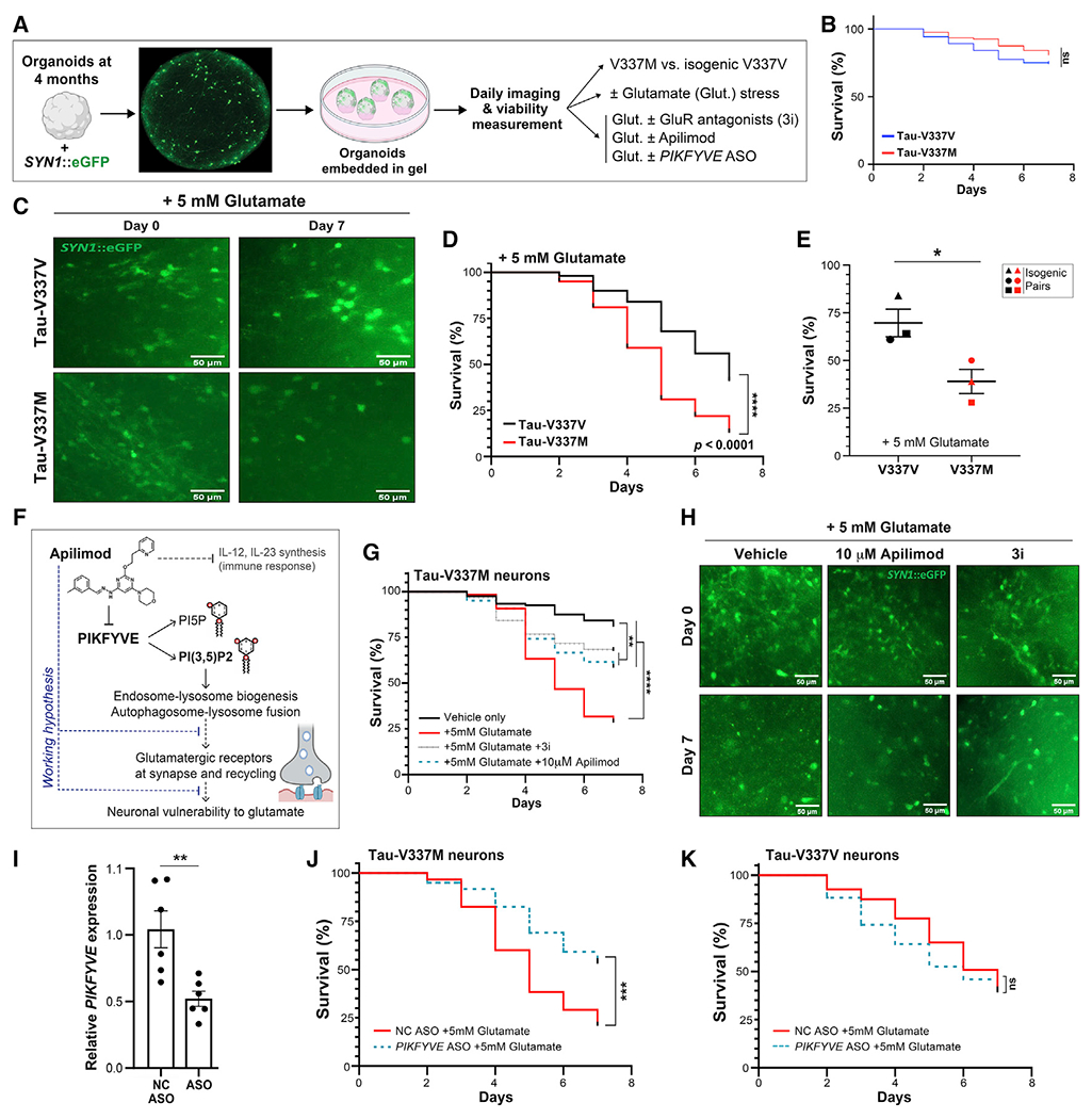
Frontotemporal dementia (FTD) because of MAPT mutation causes pathological accumulation of tau and glutamatergic cortical neuronal death by unknown mechanisms. We used human induced pluripotent stem cell (iPSC)-derived cerebral organoids expressing tau-V337M and isogenic corrected controls to discover early alterations because of the mutation that precede neurodegeneration. At 2 months, mutant organoids show upregulated expression of MAPT, glutamatergic signaling pathways, and regulators, including the RNA-binding protein ELAVL4, and increased stress granules. Over the following 4 months, mutant organoids accumulate splicing changes, disruption of autophagy function, and build-up of tau and P-tau-S396. By 6 months, tau-V337M organoids show specific loss of glutamatergic neurons as seen in individuals with FTD. Mutant neurons are susceptible to glutamate toxicity, which can be rescued pharmacologically by the PIKFYVE kinase inhibitor apilimod. Our results demonstrate a sequence of events that precede neurodegeneration, revealing molecular pathways associated with glutamate signaling as potential targets for therapeutic intervention in FTD.

Keywords: ELAVL4; MAPT; autophagy; frontotemporal dementia; glutamatergic neurons; organoids; splicing; synaptic signaling; tauopathy.


Conflict of interest statement

Declaration of interests J.D.L. employee, Amgen. A.M.G.: Scientific Advisory Board (SAB), Denali Therapeutics (2015–2018); Genetic SAB, Pfizer (2019); SAB, Genentech; consultant, GSK, AbbVie, Biogen, and Eisai. S.J.H.: SAB, Rodin Therapeutics, Frequency Therapeutics, Psy Therapeutics, Vesigen Therapeutics, and Souvien Therapeutics; inventor, patent 6,475,723. S.T.: president, StemCultures; cofounder, LUXA Biotech; SAB, Sana Biotechnology, Blue Rock Therapeutics, and Vita Therapeutics; inventor, patent 16/331,063. J.K.I.: cofounder, AcuraStem and Modulo Bio; SAB, Spinogenix. Named companies were not involved in this project.

Figures

Figure 1.
Figure 1.. Cerebral organoids exhibit similar differentiation patterns as developing fetal brains
(A) Experiment summary schematic. (B) UMAP of scRNA-seq data at 2, 4, and 6 months by cell type. Ast, astrocytes; ExDp1, excitatory deep layer 1; ExDp2, excitatory deep layer 2; ExM, maturing excitatory; ExM-U, maturing excitatory upper enriched; ExN, newborn excitatory; Glia, unspecified glia/non-neuronal cells; InCGE, interneurons caudal ganglionic eminence; InMGE, interneurons medial ganglionic eminence; IP, intermediate progenitors; OPC, oligodendrocyte precursor cells; oRG, outer radial glia; PgG2M, cycling progenitors (G2/M phase); PgS, cycling progenitors (S phase); UN, unspecified neurons; vRG, ventricular radial glia. (C) Cell type proportions (%) per line at 2, 4, and 6 months. (D) Cell type proportions (%) for individual organoids over time. Linear model, ***p < 0.001. (E) Schematic of organoid maturation and neural cell layering (left) and marker visualization (right). Confirmation of dorsal forebrain progenitors at 20 days: PAX6, SOX2, FOXG1, and Nestin; proliferation marker Ki67; absence of SOX10. 2 months: increased MAP2ab, β-III-tubulin neurons; deep-layer glutamatergic neurons TBR1, BCL11B/CTIP2; early glia S100β; few upper layer neurons SATB2+. 4–6 months: few progenitors (SOX2 and PAX6); deep- and upper-layer neurons (BRN2, MEF2C, and SATB2); GFAP+ Ast and Calbindin+ interneurons; robust tau and NeuN; expression of vGLUT1+ and pre- and post-synaptic markers SYN1 and HOMER1, respectively; white arrows indicate adjacent boutons. Scale bars, 100 μm unless otherwise indicated. See also Figure S1.
Figure 2.
Figure 2.. Tau-V337M organoids exhibit neuronal loss, early autophagy disruption, and progressive tau accumulation
(A) Proportion of glutamatergic neurons (ExDp2, ExM, and ExN) per organoid at 2, 4, and 6 months. Linear model, **p < 0.01, ***p < 0.001. (B and C) Imaging and quantification of neuronal density by NeuN+ over time in tau-V337M and isogenic V337V organoids. Mann-Whitney test, *p < 0.05. Scale bars, 250 μm (insets) and 50 μm. (D) Schematic of the ALP and key markers. (E and F) Electron photomicrographs of neurons in 2-month-old tau-V337V (E) and V337M (F) organoids. Lamellar bodies are indicated by red arrows. Scale bars, 50 μm (E) and 5 μm (F). (G–I) Western blot and densitometry quantification of ALP markers in tau-V337M and isogenic organoids at 2 (G) and 6 months (H). Relative densitometry ± SEM. Unpaired t test, *p ≤ 0.01, ***p ≤ 0.0001; n = 3 organoids per group. (J–L) Western blot and densitometry quantification of total tau and P-tau S396 levels in tau-V337M and isogenic V337V organoids at 2 (J), 4 (K), and 6 (L) months. Bars represent mean total tau densitometry or P-tau/total tau in mutant organoids (%) relative to isogenic controls (100%) ± SEM. Two-tailed unpaired t test, *p ≤ 0.01, **p ≤ 0.001, *p ≤ 0.0001; ns, non-significant; n = 9 per group from 3 independent experiments. See also Figures S2 and S3.
Figure 3.
Figure 3.. Tau-V337M organoids reveal loss of deep- and upper-layer glutamatergic neurons
(A and B) Expression of MAPT (A) and glutamatergic neuronal subtypes (ExDp1, ExDp2, ExM, and ExM-U) (B) projected onto scRNA-seq UMAPs at 2, 4, and 6 months. (C) Proportion of MAPT-expressing glutamatergic neuronal subtypes over time by mutation. Expression is scaled within each time point. Dot size, proportion of MAPT-expressing cells; color depth, MAPT expression level. Values: differential gene expression p value adjusted by model-based analysis of single-cell transcriptomics (MAST) general linear model comparisons of differential expression. (D) BCL11B/CTIP2 and MEF2C in tau-V337M (right) and isogenic V337V (left) 6-month organoids colocalized with P-tau S202/T205 (AT8) and P-tau S396/S404 (PHF1) staining. Scale bar, 10 μm. (E and F) Proportion of V337M and V337V ExDp2 neurons expressing the layer V marker BCL11B/CTIP2 (E) or ExM-U neurons expressing the layer II–IV marker MEF2C (F) at each time point; values: proportion of cells expressing each gene. MAST general linear model, *p < 0.05 **p < 0.01 ***p < 0.001 between V337M and V337V neurons. Shown are UMAPs of gene expression in ExDp2 neurons (E) or ExM-U neurons (F) at each time point. (G) Time-course image quantitative analysis of BCL11B/CTIP2+ neurons at 2,4, and 6 months normalized to DAPI. n ≥ 3 organoids from 3 separately generated organoid batches (representative image shown in D). n = number of organoids. One-way ANOVA, Tukey post hoc test, *p < 0.05, ****p < 0.0001.
Figure 4.
Figure 4.. Tau-V337M organoids exhibit early neuronal maturation and upregulation of synaptic signaling pathways
(A) Expression and connectivity of glutamatergic receptor genes and MAPT at 2 months and 2–6 months. Red, upregulation in tau-V337M organoids compared with isogenic V337V; green, downregulation; depth of color, extent of expression fold change. (B) UMAPs of glutamatergic neurons for each isogenic pair colored by mutation (top) and age (bottom). 2-month V337M-enriched clusters are indicated by black arrowheads, and 6-month V337M-enriched clusters are indicated by gray arrowheads. (C) UMAPs in (B) colored by Seurat cluster. (D and E) Z scores for enriched pathways derived from IPA for V337M-enriched 2-month (D) and 6-month (E) glutamatergic neuronal clusters (C) for each isogenic pair. (F) Network analysis constructed from significantly differentially expressed genes over time between tau-V337V and tau-V337M glutamatergic neurons following pseudobulk analysis of scRNA-seq data. Communities (C_) are labeled with ID number in bold, with the number of genes in parentheses and the most frequent parent GO term following GO enrichment and semantic similarity analysis. See also Figures S4 and S5.
Figure 5.
Figure 5.. Accelerated glutamatergic gene and ELAVL4 expression precedes aberrant splicing in V337M neurons
(A) Expression of genes with significantly differential trajectories over pseudotime in tau-V337M versus tau-V337V glutamatergic neurons by spline regression model. Trajectories grouped by unsupervised hierarchical clustering are centered across genes. Genes in significantly enriched glutamatergic signaling pathways are highlighted in cluster 2. (B) Comparison of cluster 2 pseudotime trajectories in tau-V337M and tau-V337V glutamatergic neurons. Dashed lines highlight the central region of pseudotime, where gene expression differs between mutant and control cells. (C) Trajectories of average MAPT, glutamatergic pathway gene NSG1, and ELAVL4 expression in tau-V337M and tau-V337V glutamatergic neurons over pseudotime. Adjusted p value for statistical comparison (spline regression) of trajectories is shown in the bottom right corner. (D) GO pathways enriched for DSGs between 6-month-old tau-V337M and tau-V337V organoids. (E and F) Leafcutter analysis of differentially spliced intron clusters in the glutamatergic receptor genes GRIN1 (E) and GRIA2 (F). Exons, black boxes. Red band thickness and inserted values represent proportion of spliced exon-exon pairs. (G) Expression of ELAVL4 in tau-V337M and tau-V337V glutamatergic neurons. Dot size, proportion of cells expressing ELAVL4; depth of color, ELAVL4 expression level. Values: differential gene expression p value adjusted by MAST general linear model comparisons of differential expression. (H) Number of known nELAVL gene targets by RIP (Scheckel et al., 2016) that are differentially spliced in tau-V337M organoids over time. (I) Overlap between number of DSGs in tau-V337M organoids and genes in the brain known to be bound by nELAVL by RIP analysis (Scheckel et al., 2016). (J) Semantic analysis of significant GO pathways enriched for DSGs known to be nELAVL targets (Scheckel et al., 2016). See also Figure S6.
Figure 6.
Figure 6.. ELAVL4 binds MAPT RNA and co-localizes with cytosolic stress granules in tau-V337M neurons
(A) ELAVL4 RNA immunoprecipitation (RIP) detects MAPT 3′ untranslated region (UTR) and exon 13 in tau-V337V (left panel) and tau-V337M organoids (right panel). CALM3, positive control. (B) Expression of TIA1 and G3BP1 in tau-V337M and tau-V337V glutamatergic neurons. Dot size, proportion of cells expressing a gene; depth of color, expression level. Values: differential gene expression p value adjusted by MAST general linear model. (C and D) G3BP1 immunostaining (C) and quantification of G3BP1 intensity relative to DAPI (D) in tau-V337V and V337M organoids at 2 months. Bars represent mean intensity ± SD. Unpaired t test, *p ≤ 0.01. n = 4 images per organoid; n = 3 organoids per line for 2 independent experiments. (E and F) Western blot and densitometry quantification of G3BP1 in tau-V337M and isogenic V337V organoids at 2 months. Bars represent G3BP1 densitometry in mutant organoids (%) relative to isogenic controls ± SEM. Unpaired t test, **p ≤ 0.01; n = 6 per group for 2 independent experiments. (G) ELAVL4 and G3BP1 colocalization (white arrows) in tau-V337V and V337M organoids at 2 months. Scale bar, 20 μm. (H) ELAVL4 and TIA1 co-localization with tau at 2 months (scale bar, 5 μm) and ELAVL4 with tau at 4 months (white arrows; scale bar, 10 μm). (I) Co-localization of tau, ELAVL4, and G3BP1 in tau-V337M organoids at 4 months. Scale bar, 5 μm.
Figure 7.
Figure 7.. Tau-V337M susceptibility to glutamate excitotoxicity is reversed by antagonists of excitatory receptors and PIKFYVE inhibition
(A) Longitudinal imaging method for tracking neuronal survival in cerebral organoids. (B) Survival of SYN1::GFP+ neurons in 4-month tau-V337M and isogenic V337V organoids without glutamate treatment. n = 80 neurons from 5 organoids per group. Log rank test. ns, not significant (C) Images of 4-month tau-V337M and isogenic V337V organoids treated with 5 mM glutamate. Neurons labeled with a lentivirus encoding SYN1::GFP. Scale bars, 50 μm. (D) Survival of SYN1::GFP+ neurons in tau-V337M and isogenic V337V 4-month organoids with glutamate treatment. n = 100 neurons from 5 individual organoids per group. Log rank test. ****p < 0.0001. (E) Percentage of surviving neurons following 7 days of glutamate treatment from 3- to 4-month organoids. Each point represents an independent experiment among three isogenic pairs. Two-tailed unpaired t test, *p = 0.0436. (F) Schematic of the proposed mode of action for apilimod and effect on neuronal vulnerability to excitotoxic stress. (G) Survival of SYN1::GFP+ neurons in tau-V337M organoids with glutamate treatment and DMSO. 3i, 10 μM CNQX + 10 μM MK-801 + 2 mM nimodipine or 10 μM apilimod. n = 120 neurons from 5 individual organoids per group. Log rank test, **p < 0.01 ****p < 0.0001. (H) Images of 4-month tau-V337M organoids treated with 5 mM glutamate and DMSO. 3i, 10 μM CNQX + 10 μM MK-801 + 2 μM nimodipine or 10 μM apilimod. Neurons were labeled with SYN1::GFP. Scale bars, 50 μm. (I) Relative PIKFYVE expression in ND03231 organoids by qRT-PCR following treatment with a PIKFYVE ASO, normalized to 18S expression. Bars represent mean expression ± SEM with n = 6 organoids per group. Two-tailed unpaired t test, **p < 0.01. (J and K) Survival of SYN1::GFP+ neurons in 4-month tau-V337M (J) and tau-V337V (K) organoids with glutamate and 10 μM negative control (NC) or PIKFYVE ASO treatment. n = 120 neurons from 5 individual organoids per group. Log rank test, ***p = 0.001, ****p < 0.0001. See also Figure S7.

Comment in

References

    1. Akamatsu W, Fujihara H, Mitsuhashi T, Yano M, Shibata S, Hayakawa Y, Okano HJ, Sakakibara S, Takano H,Takano T, et al. (2005). The RNA-binding protein HuD regulates neuronal cell identity and maturation. Proc. Natl. Acad. Sci. USA 102, 4625–4630. - PMC - PubMed
    1. Anderson P, and Kedersha N (2008). Stress granules: the Tao of RNA triage. Trends Biochem. Sci 33, 141–150. - PubMed
    1. Apicco DJ, Zhang C, Maziuk B, Jiang L, Ballance HI, Boudeau S, Ung C, Li H, and Wolozin B (2019). Dysregulation of RNA Splicing in Tauopathies. Cell Rep. 29, 4377–4388.e4. - PMC - PubMed
    1. Aran D, Looney AP, Liu L, Wu E, Fong V, Hsu A, Chak S, Naikawadi RP, Wolters PJ, Abate AR, et al. (2019). Reference-based analysis of lung single-cell sequencing reveals a transitional profibrotic macrophage. Nat. Immunol 20, 163–172. - PMC - PubMed
    1. Arendt T (2002). Dysregulation of neuronal differentiation and cell cycle control in Alzheimer’s disease. J. Neural Transm Suppl., 77–85. - PubMed

Publication types

MeSH terms