A comparison of subjective and objective conjunctival hyperaemia grading with AOS® Anterior software
- PMID: 34315357
- PMCID: PMC8792102
- DOI: 10.1080/08164622.2021.1945406
A comparison of subjective and objective conjunctival hyperaemia grading with AOS® Anterior software
Abstract
Clinical relevance: This study evaluates a commercially available conjunctival hyperaemia grading system, providing validation of an important tool for ocular surface research and clinical trials.
Background: Bulbar conjunctival hyperaemia is a sign of ocular surface inflammation, and proper measurement is essential to clinical care and trials. The aim of this study was to assess the validity and repeatability of an objective grading system in comparison with subjective grading.
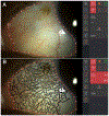
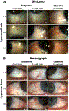
Methods: This study was a retrospective, randomised analysis of 300 bulbar conjunctival images that were collected at an academic institution. The images used were de-identified and collected from the Keratograph K5 and Haag-Streit slitlamp. Six investigators graded the images with either a 0.1 or 0.5 unit scaling using a 0-4 Efron grading scale. Three of the investigators also imported the images into the AOS ® Anterior software and graded them objectively. All measurement techniques were assessed for repeatability and comparability to each other.
Results: Mean hyperaemia with the objective system (1.1 ± 0.7) was significantly less than the subjective grading (2.0 ± 0.8) (P < 0.001). Both inter- and intra-subject repeatability of the objective system (0.15) was better than the subjective methods (1.70).
Conclusion: The results showed excellent repeatability of the AOS ® Anterior objective conjunctival hyperaemia grading software, although they were not found to be interchangeable with subjective scores. This system has value in monitoring levels of hyperaemia in contact lens wearers and patients in clinical care and research trials.
Keywords: Hyperaemia; Objective grading; Subjective grading; conjunctiva.
Figures


References
-
- Terry R, Schnider C, Holden B, et al. CCLRU standards for success of daily and extended wear contact lenses. Optom Vis Sci. 1993;70:234–43. - PubMed
-
- Efron N, Pritchard N, Brandon K, et al. A survey of the use of grading scales for contact lens complications in optometric practice. Clin Exp Optom. 2011;94:193–9. - PubMed
-
- Kurita J, Shoji J, Inada N, et al. Clinical Severity Classification using Automated Conjunctival Hyperemia Analysis Software in Patients with Superior Limbic Keratoconjunctivitis. Curr Eye Res. 2018;43:679–82. - PubMed
Publication types
MeSH terms
Grants and funding
LinkOut - more resources
Full Text Sources
Medical
Research Materials
