. 2020 Sep 25;2(3):zcaa022.
doi: 10.1093/narcan/zcaa022. eCollection 2020 Sep.

Replication protein A: a multifunctional protein with roles in DNA replication, repair and beyond

Affiliations

Replication protein A: a multifunctional protein with roles in DNA replication, repair and beyond

Rositsa Dueva et al. NAR Cancer. .

Abstract

Single-stranded DNA (ssDNA) forms continuously during DNA replication and is an important intermediate during recombination-mediated repair of damaged DNA. Replication protein A (RPA) is the major eukaryotic ssDNA-binding protein. As such, RPA protects the transiently formed ssDNA from nucleolytic degradation and serves as a physical platform for the recruitment of DNA damage response factors. Prominent and well-studied RPA-interacting partners are the tumor suppressor protein p53, the RAD51 recombinase and the ATR-interacting proteins ATRIP and ETAA1. RPA interactions are also documented with the helicases BLM, WRN and SMARCAL1/HARP, as well as the nucleotide excision repair proteins XPA, XPG and XPF-ERCC1. Besides its well-studied roles in DNA replication (restart) and repair, accumulating evidence shows that RPA is engaged in DNA activities in a broader biological context, including nucleosome assembly on nascent chromatin, regulation of gene expression, telomere maintenance and numerous other aspects of nucleic acid metabolism. In addition, novel RPA inhibitors show promising effects in cancer treatment, as single agents or in combination with chemotherapeutics. Since the biochemical properties of RPA and its roles in DNA repair have been extensively reviewed, here we focus on recent discoveries describing several non-canonical functions.


Figures

Figure 1.
Figure 1.
Schematic representation of RPA domains. (A) The domains in each subunit of RPA complex are joined by flexible linkers. RPA has four ssDNA-binding domains with DBD-A and DBD-B being high-affinity ssDNA-binding domains, as indicated by intensity gradient in ssDNA. The N-terminal domain of RPA1 (DBD-F) is involved in protein–protein interactions including tumor suppressor p53. Zinc finger motif in the C-terminal fold of 70 kDa subunit provides structural stability and has a positive role in RPA’s DNA-binding activity. The phosphorylation motif is located in the N-terminus of RPA2. RPA32C contains a winged helix–turn–helix (WH) fold involved in protein–protein interactions. Triple arrow represents the inter-subunit interactions, known as the RPA trimerization core. Two-headed arrows represent protein–protein interactions. (B) Final stage of RPA binding to ssDNA of around 30 nt. Upon DNA damage, RPA gains several negative charges through phosphorylation, primarily on the N-terminal domain of RPA2 (32N), which alters RPA conformation and induces its physical interaction with the N-terminus of RPA1 (128). Electrostatic repulsive forces between hyperphosphorylated RPA2 and negatively charged ssDNA may foster RPA dissociation from ssDNA.
Figure 2.
Figure 2.
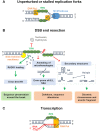
RPA binding to ssDNA intermediates. (A) RPA binds to ssDNA intermediates during DNA synthesis under normal conditions and when replication forks are stalled by genotoxic agents. (B) DNA end resection also creates ssDNA, which is substrate for all forms of homology-directed repair (HDR). RPA has a protective role against nucleases and formation of secondary structures such as G-quadruplexes (G4s) and hairpins, thus supporting RAD51-mediated HR. Furthermore, RPA prevents spontaneous annealing of microhomologies and inverted repeats that can lead to deletions, sequence alterations or chromosome breakage with the accompanying dicentric chromosomes and acentric fragments. (C) The displaced ssDNA strand during transcription can be recognized by RPA in the absence of DNA damage. Note that other components involved in these processes are not shown for simplicity.
Figure 3.
Figure 3.
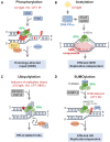
RPA modifications coordinate DNA repair. RPA’s modifications vary between the genotoxic agents and the cell cycle phase. (A) RPA is phosphorylated during normal cell cycle by CDKs and hyperphosphorylated at multiple serine/threonine residues by PIKK kinases in response to genotoxic stress. (B) UV light is the trigger for RPA acetylation primarily at a single lysine residue. (C) ssDNA also causes RPA ubiquitylation at multiple lysines. (D) Association between RPA1 and SENP6 during S phase keeps RPA70 in a hypo-SUMOylated state. Inducers of genotoxic stress are indicated in deep red. Note that other components involved in these processes are not shown for simplicity.
Figure 4.
Figure 4.
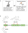
Clinical applications of RPA. (A) Overexpression of RPA subunits or hyperphosphorylation of RPA2 may serve as a prognostic biomarker in tumor specimens. This fact guided the development of novel RPA inhibitors, which have the potential to inhibit DNA replication in cancer cells. (B) Examples of well-characterized small molecule inhibitors targeting RPA1 subunit. NSC15520 (fumaropimaric acid) and HAMNO ((1Z)-1-[(2-hydroxyanilino)methylidene]naphthalen-2-one) target the N-terminal DBD-F domain, which is involved in protein–protein interactions. TDRL-505 targets the central ssDNA-binding domains of RPA1, DBD-A and DBD-B.

References

    1. Wold M.S., Kelly T. Purification and characterization of replication protein A, a cellular protein required for in vitro replication of simian virus 40 DNA. Proc. Natl Acad. Sci. U.S.A. 1988; 7:2523–2527. - PMC - PubMed
    1. Kenny M.K., Lee S.-H., Hurwitz J. Multiple functions of human single-stranded-DNA binding protein in simian virus 40 DNA replication: single-strand stabilization and stimulation of DNA polymerases α and δ. Proc. Natl Acad. Sci. U.S.A. 1989; 86:9757–9761. - PMC - PubMed
    1. Tsurimoto T., Fairman M.P., Stillman B. Simian virus 40 DNA replication in vitro: identification of multiple stages of initiation. Mol. Cell. Biol. 1989; 9:3839–3849. - PMC - PubMed
    1. Brill S.J., Stillman B. Yeast replication factor-A functions in the unwinding of the SV40 origin of DNA replication. Nature. 1989; 342:92–95. - PubMed
    1. Arunkumar A.I., Stauffer M.E., Bochkareva E., Bochkarev A., Chazin W.J. Independent and coordinated functions of replication protein A tandem high affinity single-stranded DNA binding domains. J. Biol. Chem. 2003; 278:41077–41082. - PubMed