

Fontan Liver Lesions: Not Always HCC
- PMID: 34316779
- PMCID: PMC8301494
- DOI: 10.1016/j.jaccas.2019.05.031
Fontan Liver Lesions: Not Always HCC
Abstract
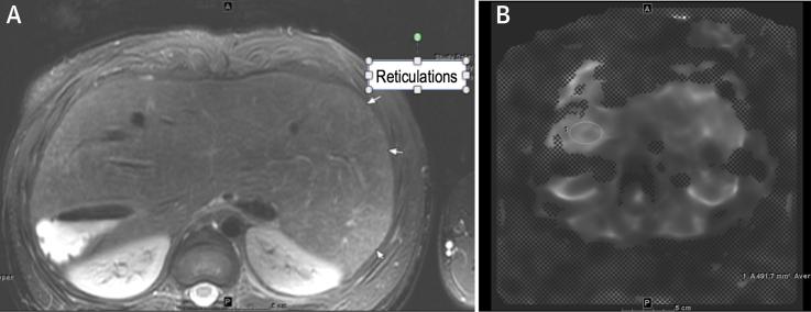
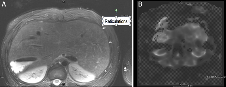
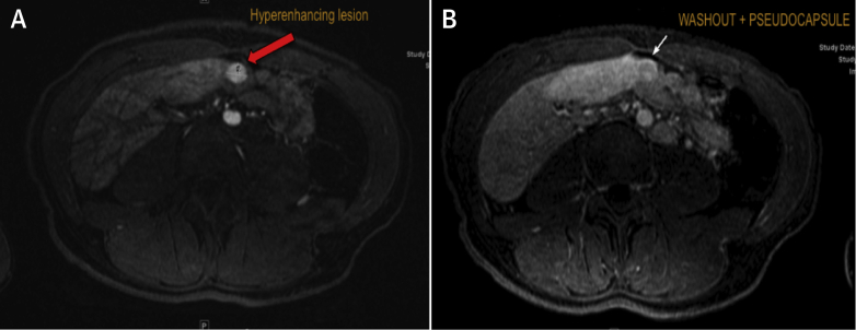
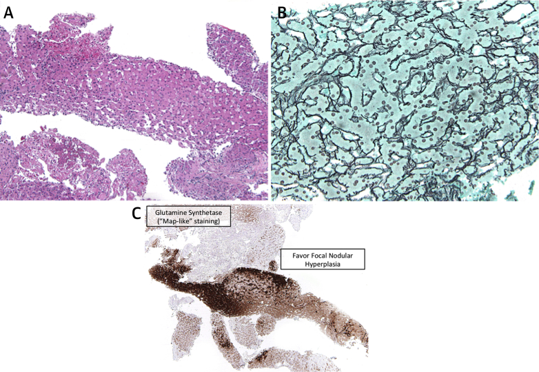
A 24-year-old Fontan procedure patient underwent surveillance liver cardiac magnetic resonance imaging. Findings were suggestive of hepatocellular carcinoma (HCC). Currently, HCC is diagnosed based on imaging alone. Given her otherwise reassuring clinical profile, she underwent liver biopsy. Pathology demonstrated focal nodular hyperplasia. This raises concern for overdiagnosis of HCC in Fontan patients without tissue confirmation. (Level of Difficulty: Advanced.).
Keywords: AASLD, American Association for Study of Liver Disease; AFP, alpha-fetoprotein; ALT, lanine aminotransferase; AST, aspartate aminotransferase; AV, atrioventricular; CT, computed tomography; FALD, Fontan associated liver disease; FNH, focal nodular hyperplasia; GS, glutamine synthetase; HCC, hepatocellular carcinoma; INR, international normalized ratio; MRI, magnetic resonance imaging; US, ultrasound; awareness; congenital heart defect; cyanotic heart disease; imaging; treatment.
© 2019 The Authors.
Figures
References
-
- Silva M.A., Hegab B., Hyde C. Needle track seeding following biopsy of liver lesions in the diagnosis of hepatocellular cancer: a systematic review and meta-analysis. Gut. 2008;57:1592–1596. - PubMed
-
- Pundi K., Pundi K.N., Kamath P.S. Liver Disease in Patients After the Fontan Operation. Am J Cardiol. 2016;117:456–460. - PubMed
-
- Asrani K., Warnes C.A., Kamath P.S. Hepatocellular Carcinoma after the Fontan Procedure. N Engl J Med. 2013;368:1756–1757. - PubMed
-
- Bryant T., Ahmad Z., Millward-Sadler H. Arterialised hepatic nodules in the Fontan circulation: hepatico-cardiac interactions. Int J Cardiol. 2011;151:268–272. - PubMed
-
- Marrero J.A., Kulik L.M., Sirlin C.B. Diagnosis, staging, and management of hepatocellular carcinoma: 2018 practice guidance by the AASLD. Hepatology. 2018;68:723. - PubMed
Publication types
LinkOut - more resources
Full Text Sources
