Case Reports
. 2020 Nov 18;2(13):2078-2084.
doi: 10.1016/j.jaccas.2020.09.042. eCollection 2020 Nov.

A Rare Case of Severe Nontropical Isolated Right Ventricular Endomyocardial Fibrosis

Affiliations
Case Reports

A Rare Case of Severe Nontropical Isolated Right Ventricular Endomyocardial Fibrosis

Shaaheen Laher et al. JACC Case Rep. .

Abstract

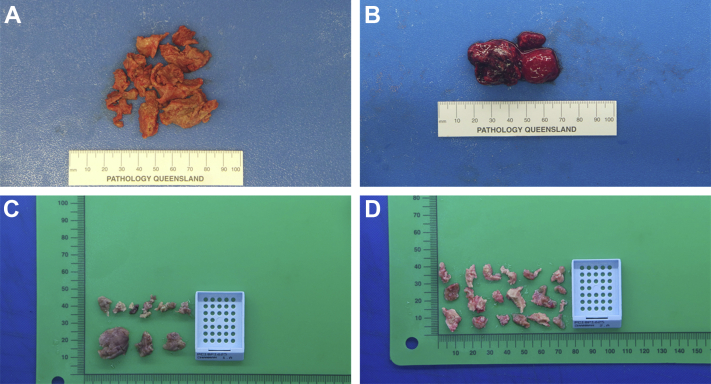
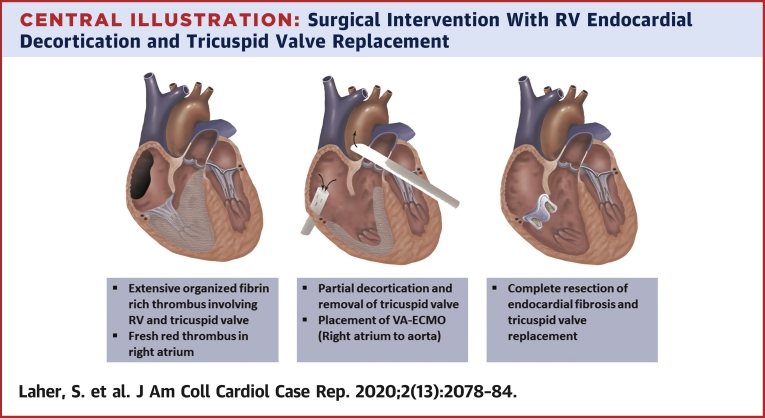
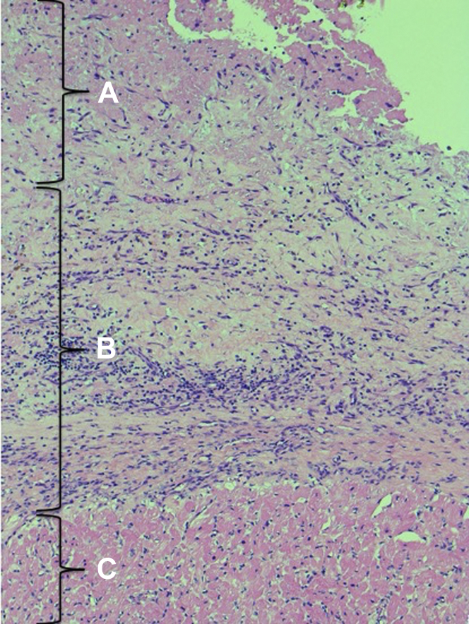
We present a case of late presentation nontropical endomyocardial fibrosis isolated to the right ventricle and tricuspid valve (TV). In response to deteriorating hemodynamics, surgical debulking and TV removal were performed before initiation of centralized venoarterial extracorporeal membrane oxygenation support. Definitive endomyocardial resection with a TV prosthesis was then successfully completed. (Level of Difficulty: Advanced.).

Keywords: CE, contrast-enhanced; CMR, cardiac magnetic resonance; ECMO, extracorporeal membrane oxygenation; EMF, endomyocardial fibrosis; LV, left ventricular; RA, right atrial; RV, right ventricular; RVOT, right ventricular outflow tract; TTE, Transthoracic echocardiogram; TV, tricuspid valve; contrast enhanced TTE; endomyocardial fibrosis; right ventricle.


Conflict of interest statement

Dr. Platts is acting as a medical liaison for Lantheus Medical Imaging. All other authors have reported that they have no relationships relevant to the contents of this paper to disclose.

Figures

None
Graphical abstract
Figure 1
Figure 1
Transthoracic Echocardiography on Presentation (A) Apical 4-chamber view showing obliteration of the right ventricular (RV) cavity (upper arrow) and an echogenic mass in the right atrial (RA) appendage (lower arrow). (B) Parasternal long-axis view showing echogenic mass filling the right ventricular cavity (upper arrow) with mitral valve prolapse (lower arrow). (C) Parasternal short-axis view showing the outline of the right ventricular cavity. (D) Midsystolic color Doppler imaging demonstrating severe tricuspid regurgitation (arrow). LV = left ventricle.
Figure 2
Figure 2
Contrast-Enhanced Transthoracic Echocardiography (A to D) Contrast-enhanced transthoracic echocardiography showing that the right ventricular cavity is nearly obliterated by a homogenous echo-dense mass. The mass displays well-defined endocardial tissue planes. The use of very low mechanical index myocardial contrast echocardiography with flash destruction replenishment sequences reveals abnormal myocardial perfusion and an avascular mass consistent with thrombus.
Figure 3
Figure 3
Cardiac Magnetic Resonance Imaging (A) A 4-chamber view demonstrating a dilated right ventricle with impaired contraction and cavity obliteration (blue arrow) and large bilateral pleural effusions (white arrows). (B) Right ventricular outflow tract view demonstrating obliteration of the right ventricular cavity (blue arrow) and pleural effusion (white arrow). (C) Immediate post–gadolinium enhancement image with large thrombi in the right atrial appendage (white arrowhead) and filling the right ventricular apex (blue arrow). (D) Late gadolinium enhancement image in the same plane as C and showing a triangular thrombus in the right ventricular apex (blue arrow) surrounded by bright subendocardial enhancement (blue arrowheads), confirming the diagnosis of endomyocardial fibrosis.
Figure 4
Figure 4
Surgical Resection Images obtained during endocardial decortication.
Figure 5
Figure 5
Endocardial Decortication (A, C, D) The right ventricle was infiltrated with chronic, organized, and fibrin-rich thrombus. (B) The right atrium had evidence of fresh, red thrombus secondary to stasis.
Central Illustration
Surgical Intervention With RV Endocardial Decortication and Tricuspid Valve Replacement RV = right ventricular; VA-ECMO = venoarterial-extracorporeal membrane oxygenation.
Figure 6
Figure 6
Endocardial fibrosis With Neovascularization, Mild Mixed Inflammatory Cell Infiltrate and Organizing Surface Fibrin Hematoxylin and Eosin, ×100) The slide reveals a surface layer of fibrin (A) and neovascularized endocardium with fibrosis and mild mixed inflammatory cell infiltrate in the middle (B). The bottom layer shows myocardium (C).

References

    1. Grimaldi A., Mocumbi A.O., Freers J. Tropical endomyocardial fibrosis: natural history, challenges, and perspectives. Circulation. 2016;133:2503–2515. - PubMed
    1. Bukhman G., Ziegler J., Parry E. Endomyocardial fibrosis: still a mystery after 60 years. PLoS Negl Trop Dis. 2008;2:e97. - PMC - PubMed
    1. Mankad R., Bonnichsen C., Mankad S. Hypereosinophilic syndrome: cardiac diagnosis and management. Heart. 2016;102:100–106. - PubMed
    1. Mocumbi A.O., Ferreira M.B., Sidi D., Yacoub M.H. A population study of endomyocardial fibrosis in a rural area of Mozambique. N Engl J Med. 2008;359:43–49. - PubMed
    1. Moraes F., Lapa C., Hazin S., Tenorio E., Gomes C., Moraes C.R. Surgery for endomyocardial fibrosis revisited. Eur J Cardiothorac Surg. 1999;15:309–312. discussion 312–3. - PubMed

Publication types

LinkOut - more resources