Case Reports
. 2020 Oct 14;2(14):2156-2161.
doi: 10.1016/j.jaccas.2020.07.056. eCollection 2020 Nov 18.

Unusual Cause of Severe Tricuspid Regurgitation: Tricuspid Leaflet Annular Tear Following Remote Motor Vehicle Accident

Affiliations
Case Reports

Unusual Cause of Severe Tricuspid Regurgitation: Tricuspid Leaflet Annular Tear Following Remote Motor Vehicle Accident

Daniel G Bamira et al. JACC Case Rep. .

Abstract

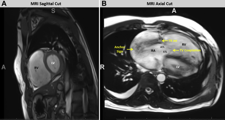
Tricuspid regurgitation (TR) is an uncommon and underdiagnosed complication of blunt chest trauma. Typical mechanisms include torn chordae, papillary muscle rupture, and radial leaflet tear. We describe an unusual case of traumatic TR due to circumferential avulsion of the anterior tricuspid leaflet from the tricuspid annulus and the crucial role of multimodality imaging in its diagnosis and treatment. (Level of Difficulty: Intermediate.).

Keywords: 3D, 3-dimensional; CT, computed tomography; RA, right atrium; RV, right ventricle; TR, tricuspid regurgitation; TV, tricuspid valve; avulsion; cardiac magnetic resonance imaging; leaflet tear; motor vehicle accident; transesophageal echocardiography; tricuspid valve.


Conflict of interest statement

The authors have reported that they have no relationships relevant to the contents of this paper to disclose.

Figures

None
Graphical abstract
Figure 1
Figure 1
2-Dimensional Transesophageal Echocardiogram Focused on Right Heart Demonstrates Avulsion of Anterior Leaflet of the Tricuspid Valve on Gray-Scale Imaging Resulting in Severe Eccentric TR 2-dimensional transesophageal echocardiogram focused on the right heart demonstrates avulsion of anterior leaflet of the tricuspid valve on gray-scale imaging (A), resulting in severe eccentric tricuspid regurgitation (TR) (B). (Inset) Spectral Doppler of the TR demonstrates dense, triangular shaped, low-velocity jet, consistent with severe TR. ATL = anterior tricuspid leaflet; LA = left atrium; LV = left ventricle; RA = right atrium; RV = right ventricle.
Figure 2
Figure 2
2-Dimensional Transesophageal Echocardiogram Focused on Right Heart Shows the TR Jet Extending to the Posterior RA Wall Giving Rise to the So-Called Anchor Sign Abbreviations as in Figure 1.
Figure 3
Figure 3
2-Dimensional Transesophageal Echocardiogram Delineating the Tricuspid Leaflet Coaptation Point Versus the Site of Avulsion 2-dimensional transesophageal echocardiogram delineating the tricuspid leaflet coaptation point versus the site of avulsion (A) with severe TR at the site of valve avulsion versus mild central TR (B). ATL = anterior tricuspid leaflet; CS = coronary sinus; other abbreviations as in Figure 1.
Figure 4
Figure 4
3-Dimensional Transesophageal Echocardiogram Imaging Shows Circumferential Avulsion of the ATL From the Tricuspid Annulus 3-dimensional transesophageal echocardiogram (3D TEE) imaging demonstrating circumferential avulsion of the anterior tricuspid leaflet from the tricuspid annulus as visualized from the RA on standard (A) and photorealistic TrueVUE rendering (TrueVUE, Sewell, New Jersey) (B). Asterisk and arrow indicate the orifice resulting from avulsion. AV = aortic valve; other abbreviations as in Figures 1 and 3.
Figure 5
Figure 5
3-Dimensional Transesophageal Echocardiogram Imaging Demonstrating Circumferential Avulsion of the ATL 3-dimensional transesophageal echocardiogram imaging shows circumferential avulsion of the anterior tricuspid leaflet from the tricuspid annulus as visualized from the RV on standard (A) and photorealistic TrueVue rendering (B). Asterisk and arrow point to the orifice resulting from the avulsion. Abbreviations as in Figures 1, 3, and 4.
Figure 6
Figure 6
Chest CT Reveals Markedly Dilated Right Heart With Contrast Reflux Chest computed tomography (CT) reveals markedly dilated right heart (A, axial cut) with contrast reflux into a dilated inferior vena cava and hepatic veins (B, coronal cut), consistent with chronic severe TR. IVC = inferior vena cava; other abbreviations as in Figure 1.
Figure 7
Figure 7
Cardiac Magnetic Resonance Shows a Dilated RV and TR Cardiac magnetic resonance shows a dilated RV (A) and TR originating along the base of the anterior leaflet adjacent to the TV annular hinge point and distinct from the milder transvalvular TR (B). ATL = anterior tricuspid leaflet; STL = septal tricuspid leaflet; other abbreviations as in Figure 1.
Figure 8
Figure 8
Intraoperative Views of the Opened Right Atrium Showing the Avulsion of the ATL Intraoperative views of the opened right atrium showing the avulsion of the ATL from the TV annulus (A) and the relationship between the ATL and other tricuspid leaflets. (B) PTL = posterior tricuspid leaflet; RAA = right atrial appendage; STL = septal tricuspid leaflet; other abbreviations as in Figure 1.

References

    1. Avegliano G., Corneli M., Conde D., Ronderos R. Traumatic rupture of the tricuspid valve and multi-modality imaging. Cardiovasc Diagn Ther. 2014;4:401–405. - PMC - PubMed
    1. Mehrotra D., Dalley P., Mahon B. Tricuspid valve avulsion after blunt chest trauma. Tex Heart Inst J. 2012;39:668–670. - PMC - PubMed
    1. Gayet C., Pierre B., Delahaye J.P., Champsaur G., Andre-Fouet X., Rueff P. Traumatic tricuspid insufficiency. An underdiagnosed disease. Chest. 1987;92:429–432. - PubMed
    1. Kulik A., Al-Saigh M., Yelle J.D., Rubens F.D. Subacute tricuspid valve rupture after traumatic cardiac and pulmonary contusions. Ann Thorac Surg. 2006;81:1111–1112. - PubMed
    1. Williams A. Laceration of the tricuspid valve. London Medical Gazette. 1829:78–79.

Publication types

LinkOut - more resources