Case Reports
. 2020 Dec 16;2(15):2304-2309.
doi: 10.1016/j.jaccas.2020.09.032. eCollection 2020 Dec.

An Uncommon Cause of ST-Segment Elevation Myocardial Infarction: Intramural Coronary Artery Hematoma After Blunt Chest Trauma

Affiliations
Case Reports

An Uncommon Cause of ST-Segment Elevation Myocardial Infarction: Intramural Coronary Artery Hematoma After Blunt Chest Trauma

Diego H González-Bravo et al. JACC Case Rep. .

Abstract

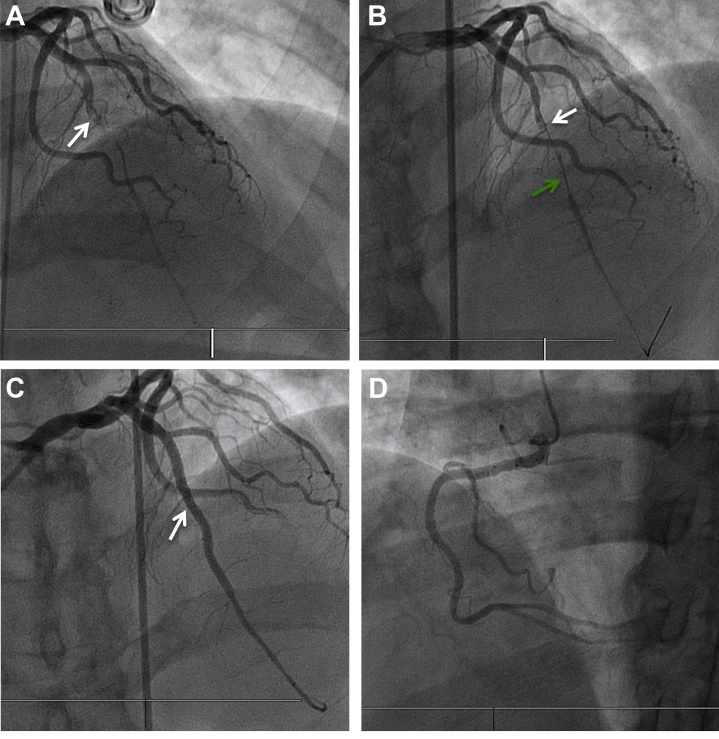
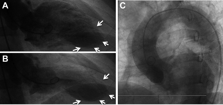
Intramural coronary hematoma (IMCH) is a rare cause of acute myocardial infarction (MI). We aim to review the current knowledge and share our experience with the diagnosis and management of a patient presenting with traumatic IMCH leading to an acute ST-segment elevation MI. (Level of Difficulty: Intermediate.).

Keywords: BCT, blunt chest trauma; CP, chest pain; DAPT, dual anti-platelet therapy; ECG, electrocardiogram; IMCH, intramural coronary hematoma; IVUS, intravascular ultrasound; LAD, left anterior descending artery; MI, myocardial infarction; PCI, percutaneous coronary intervention; SCAD, spontaneous coronary artery dissection; STEMI, ST segment elevation myocardial infarction; TIMI, Thrombolysis In Myocardial Infarction; acute coronary syndrome; chest pain; dissection; intravascular ultrasound; myocardial infarction; percutaneous coronary intervention.


Conflict of interest statement

This material is the result of work supported with resources and the use of facilities at the Veterans Affairs, Caribbean Healthcare System San Juan, P.R. The contents of this publication do not represent the views of the Veterans Affairs Caribbean Healthcare System, the U.S. Department of Veterans Affairs, or the United States Government. All authors have reported that they have no relationships relevant to the contents of this paper to disclose.

Figures

None
Graphical abstract
Figure 1
Figure 1
Initial Electrocardiogram Initial electrocardiogram with sinus rhythm and ST-segment elevation (red arrows) in leads V2 to V6 with associated Q waves suggestive of anterolateral ST-segment elevation myocardial infarction.
Figure 2
Figure 2
Chest X-Ray Chest X-ray without acute cardiopulmonary disease and no evident bone fractures.
Figure 3
Figure 3
Coronary Angiography (A) Subtotal occlusion of mid left anterior descending artery with Thrombolysis In Myocardial Infarction flow grade 1 and filling defect suggestive of thrombus (white arrow) (Video 1). (B) After performing aspiration thrombectomy to the mid left anterior descending artery (white arrow) and providing 200 μg of intracoronary nitroglycerin, a long and smooth tubular lesion persisted (green arrow), which raised suspicion for external compression given the history of recent blunt chest trauma. (C) Final result (Video 3) after intravascular ultrasound-guided stenting of the mid left anterior descending artery (white arrow). (D) Normal right coronary artery.
Figure 4
Figure 4
Intravascular Ultrasound (A to C) Intravascular ultrasound frames showing extensive spiral intramural hematoma (∗) of the mid to distal left anterior descending artery with lumen compromise (Video 2). (D) After Intravascular ultrasound-guided stenting of mid left anterior descending artery with compressed residual intramural hematoma (arrow).
Figure 5
Figure 5
Ventricular and Aortic Angiography Ventricular diastolic (A) and systolic (B) frames with evidence of distal inferoapical and anteroapical akinesis (white arrows) corresponding with the wrap-around left anterior descending artery territory and mildly reduced overall systolic function (ejection fraction 40% to 45%). (C) Ascending aorta angiography without evidence of dissection or aneurysm
Figure 6
Figure 6
Transthoracic Echocardiogram Post-Myocardial Infarction Day 1 Two-chamber diastolic (A) and systolic (B) frames showing severe hypokinesis of antero-apical, infero-apical, and mid anteroseptal walls. Four-chamber diastolic (C) and systolic (D) frames with intravenous contrast showing severe hypokinesis of the septal and lateral apical walls. No evidence of left ventricular thrombi, pericardial effusion, or major valvulopathies.

References

    1. James M.M., Verhofste M., Franklin C., Beilman G., Goldman C. Dissection of the left main coronary artery after blunt thoracic trauma: case report and literature review. World J Emerg Surg. 2010;5:21. - PMC - PubMed
    1. Lempereur M., Fung A., Saw J. Stent mal-apposition with resorption of intramural hematoma with spontaneous coronary artery dissection. Cardiovasc Diagn Ther. 2015;5:323–329. - PMC - PubMed
    1. Moidy M., Al Kindi F. Coronary intramural hematoma: challenges in diagnosis and management. Heart Views. 2019;20:17–20. - PMC - PubMed
    1. Shenoda M. Aragon J. Coronary intramural hematomas: a focused review. Interv Cardiol. 2015;7:239–249.
    1. Christensen M.D., Nielsen P.E., Sleight P. Prior blunt chest trauma may be a cause of single vessel coronary disease; hypothesis and review. Int J Cardiol. 2006;108:1–5. - PubMed

Publication types

LinkOut - more resources