

Case Reports
doi: 10.1016/j.jaccas.2019.11.032.
eCollection 2020 Feb.
"Double-Parachute" Mitral Valve
Affiliations
- PMID: 34317218
- PMCID: PMC8298313
- DOI: 10.1016/j.jaccas.2019.11.032
Item in Clipboard
Case Reports
"Double-Parachute" Mitral Valve
JACC Case Rep.
.
Abstract
A 38-year-old asymptomatic man was referred by his general practitioner for a 3/6 systolic heart murmur, which was detected during a routine consultation. Echocardiography revealed a parachute mitral valve associated with a parachute-like membrane, causing significant subaortic obstruction that was eventually surgically resected with an excellent postoperative outcome. (Level of Difficulty: Beginner.).
Keywords: AMVT, accessory mitral valve tissue; LVOT, left ventricular outflow tract; MR sequences; MRI, magnetic resonance imaging; PMV, parachute mitral valve; congenital heart disease; echocardiography; mitral valve.
© 2020 The Authors.
Figures


Transthoracic Echocardiography (A) Accessory mitral valve tissue (AMVT) protruding through the aortic valve, parasternal long-axis view. (B) AMVT attached to the anterior mitral leaflet (AML), parasternal short-axis view. (C) Single posteromedial papillary muscle (PM), parasternal short-axis view. (D) High velocities in the left ventricular outflow tract measured by continuous wave Doppler (Pedoff transducer), nonstandard right parasternal view. See Video 1.
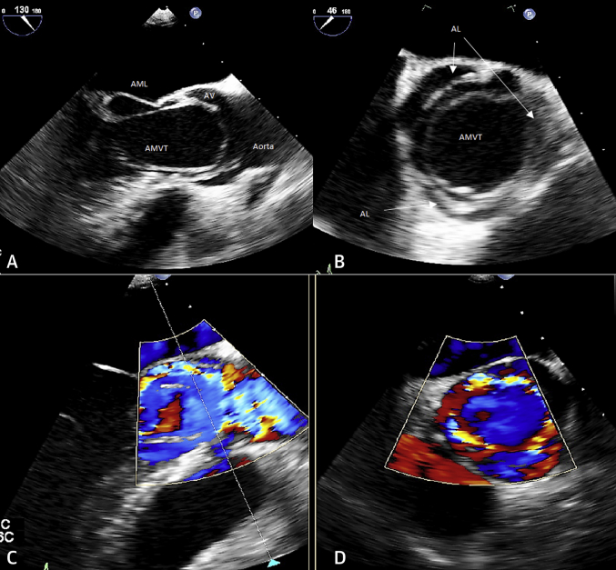
Transesophageal Echocardiography (A) AVMT membrane protruding through the AV and attached to the AML, mid-esophageal long-axis view. (B) AVMT membrane protruding between opened AL, mid-esophageal short-axis view. (C) Anterograde aortic flow around the AVMT membrane on color Doppler, mid-esophageal long-axis view. (D) Anterograde aortic flow around AVMT membrane on color Doppler, mid-esophageal short-axis view. See Videos 2 and 3. AL = aortal valve leaflets; AV = aortic valve; other abbreviations as in Figure 1.

Cardiac MRI AMVT attached to both the AML and the posteromedial PM on a long-axis view, cine cardiac MRI images. See Video 4. LA = left atrium; LV = left ventricle; MRI = magnetic resonance imaging; other abbreviations as in Figure 1.
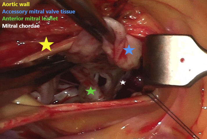
Surgical View of the Accessory Mitral Valve Tissue
References
-
- Oosthoek P.W., Wenink A.C., Wisse L.J., Gittenberger-de Groot A.C. Development of the papillary muscles of the mitral valve: morphogenetic background of parachute-like asymmetric mitral valves and other mitral valve anomalies. J Thorac Cardiovasc Surg. 1998;116:36–46. - PubMed
-
- Shone J.D., Sellers R.D., Anderson R.C., Adams P., Jr., Lillehei C.W., Edwards J.E. The developmental complex of “parachute mitral valve,” supravalvular ring of left atrium, subaortic stenosis, and coarctation of aorta. Am J Cardiol. 1963;11:714–725. - PubMed
-
- Hakim F.A., Kendall C.B., Alharthi M., Mancina J.C., Tajik J.A., Mookadam F. Parachute mitral valve in adults-a systematic overview. Echocardiography. 2010;27:581–586. - PubMed
-
- Stout K.K., Daniels C.J., Aboulhosn J.A. 2018 AHA/ACC guideline for the management of adults with congenital heart disease: a report of the American College of Cardiology/American Heart Association task force on clinical practice guidelines. J Am Coll Cardiol. 2019;73:e81–e192. - PubMed
-
- Prifti E., Bonacchi M., Bartolozzi F., Frati G., Leacche M., Vanini V. Postoperative outcome in patients with accessory mitral valve tissue. Med Sci Monit. 2003;9:RA126–RA133. - PubMed
Publication types
LinkOut - more resources
Full Text Sources
