

An Exceptional Cause of Acute Right Heart Failure: Isolated Right Ventricular Takotsubo Syndrome
- PMID: 34317243
- PMCID: PMC8311621
- DOI: 10.1016/j.jaccas.2019.10.038
An Exceptional Cause of Acute Right Heart Failure: Isolated Right Ventricular Takotsubo Syndrome
Abstract
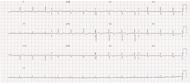
We describe a patient with of acute right ventricular dysfunction secondary to right ventricular isolated Takotsubo syndrome (TTS). The importance of appropriate differential diagnosis for acute right ventricular dysfunction differential diagnosis of acute right ventricular dysfunction and the differences in diagnosis and management of right ventricular TTS and typical left ventricular TTS are highlighted. (Level of Difficulty: Intermediate.).
Keywords: AMI, acute myocardial infarction; ECG, electrocardiogram; LV, left ventricle; MRI, magnetic resonance imaging; RV, right ventricle; TTS, Takotsubo syndrome; Takotsubo syndrome; cardiac magnetic resonance; cardiogenic shock; right ventricle; stress cardiomyopathy.
© 2020 The Authors.
Figures


Comment in
-
The Ever-Expanding Landscape of Cardiomyopathies.JACC Case Rep. 2020 Mar 18;2(3):361-364. doi: 10.1016/j.jaccas.2020.02.006. eCollection 2020 Mar. JACC Case Rep. 2020. PMID: 34319296 Free PMC article.
References
-
- Liu K., Carhart R. “Reverse McConnell’s sign?”: a unique right ventricular feature of Takotsubo cardiomyopathy. Am J Cardiol. 2013;111:1232–1235. - PubMed
-
- Lyon A.R., Bossone E., Schneider B. Current state of knowledge on Takotsubo syndrome: a position statement from the Taskforce on Takotsubo Syndrome of the Heart Failure Association of the European Society of Cardiology. Eur J Heart Fail. 2016;18:8–27. - PubMed
-
- Stähli B.E., Ruschitzka F., Enseleit F. Isolated right ventricular ballooning syndrome: a new variant of transient cardiomyopathy. Eur Heart J. 2011;32:1821. - PubMed
Publication types
LinkOut - more resources
Full Text Sources
