Case Reports
. 2020 Feb 12;2(3):378-383.
doi: 10.1016/j.jaccas.2019.11.072. eCollection 2020 Mar.

Cardiac Myxoma in a Patient With Hypertrophic Cardiomyopathy

Affiliations
Case Reports

Cardiac Myxoma in a Patient With Hypertrophic Cardiomyopathy

Weng-Tein Gi et al. JACC Case Rep. .

Abstract

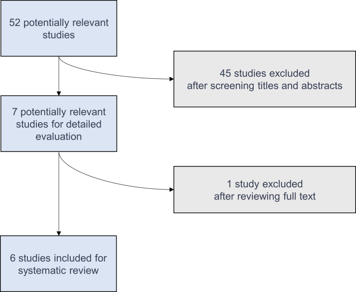
We report a rare case of concomitant hypertrophic cardiomyopathy and cardiac myxoma without LEOPARD syndrome. Additionally, 6 similar cases were systemically reviewed, and the characteristics of this first-ever studied patient group were summarized. (Level of Difficulty: Beginner.).

Keywords: ECG, electrocardiogram; FO, fossa ovalis; HCM, hypertrophic cardiomyopathy; LEOPARD syndrome; LGE, late gadolinium enhancement; LVOT, left ventricular outflow tract; NT-proBNP, N-terminal pro–B-type natriuretic peptide; SCD, sudden cardiac death; cardiac myxoma; echocardiography; hs-cTnT, high-sensitivity cardiac troponin T; hypertrophic cardiomyopathy; imaging.


Figures

None
Graphical abstract
Figure 1
Figure 1
Electrocardiography and Transthoracic Echocardiogram (A) A 12-lead ECG shows giant T-wave inversions and ST-segment depressions in the precordial leads. (B) Transthoracic echocardiography shows a hypertrophied LV with a thickened interventricular septum (red arrow). (C) A broad-based mass (dotted red arrow) is shown attached to the interatrial septum in the LA. ECG = electrocardiography; LA = left atrium; LV = left ventricle; RA = right atrium; RV = right ventricle.
Figure 2
Figure 2
Transesophageal Echocardiogram Transesophageal echocardiogram demonstrates (A) a friable multilobular tumor (solid red arrow) originating from the fossa ovalis (FO) (dotted red arrow). (B) A 3-dimensional transesophageal echocardiogram visualizes the myxoma in the left atrium. The border of the FO is denoted (dotted white line). FO = fossa ovalis; LA = left atrium; IAS = interatrial septum; RA = right atrium.
Figure 3
Figure 3
Excised Cardiac Myxoma (A) Photograph shows part of the excised cardiac myxoma (solid white arrow). Other parts of the tumor were already suctioned away during the surgery because of the tumor’s friability. The resected fossa ovalis is also shown (dashed white arrow). (B) Microscopy confirmed the features of a cardiac myxoma: an abundant myxoid stroma with inflammatory cells, small vessels, and small elongated tumor cell groups (H&E stain; ×100 original magnification). (C) Myxoid stroma with inflammatory cells and isolated siderophages (periodic acid--Schiff stain; ×200 original magnification). H&E = hematoxylin and eosin.
Figure 4
Figure 4
Cardiac Magnetic Resonance in 4-Chamber Views and Short Axis (A and B) SSFP cine sequences of the 4-chamber views and short axis respectively. The slightly hypointense tumor-occupying region (red arrow) is hardly differentiable from normal flow phenomenon. (C) Phase-sensitive inversion-recovery sequence shows diffuse intramural LGE (dotted red arrow). (D) An illustration summarizing the distribution of LGE. Ao = ascending aorta; IVS = interventricular septum; LA = left atrium; LGE = late gadolinium enhancement; LV = left ventricle; RA = right atrium; RV = right ventricle; RVOT = right ventricular outflow tract; SSFP = steady-state free precession.
Figure 5
Figure 5
Flow Chart of Studies Inspected in the Systematic Review

Comment in

References

    1. Perez de Isla L., de Castro R., Zamorano J.L. Diagnosis and treatment of cardiac myxomas by transesophageal echocardiography. Am J Cardiol. 2002;90:1419–1421. - PubMed
    1. Abbas A., Garfath-Cox K.A., Brown I.W., Shambrook J.S., Peebles C.R., Harden S.P. Cardiac MR assessment of cardiac myxomas. Br J Radiol. 2015;88:20140599. - PMC - PubMed
    1. Martinez-Quintana E., Rodriguez-Gonzalez F. LEOPARD syndrome: clinical features and gene mutations. Mol Syndromol. 2012;3:145–157. - PMC - PubMed
    1. Sala V., Gallo S., Leo C., Gatti S., Gelb B.D., Crepaldi T. Signaling to cardiac hypertrophy: insights from human and mouse RASopathies. Mol Med. 2012;18:938–947. - PMC - PubMed
    1. Elliott P.M., Anastasakis A., Borger M.A. 2014 ESC guidelines on diagnosis and management of hypertrophic cardiomyopathy: the Task Force for the Diagnosis and Management of Hypertrophic Cardiomyopathy of the European Society of Cardiology (ESC) Eur Heart J. 2014;35:2733–2779. - PubMed

Publication types