

Case Reports
doi: 10.1016/j.jaccas.2019.12.026.
eCollection 2020 Mar.
Eagle Syndrome: A Unique Cause of Carotid Bleeding
Affiliations
- PMID: 34317261
- PMCID: PMC8311603
- DOI: 10.1016/j.jaccas.2019.12.026
Item in Clipboard
Case Reports
Eagle Syndrome: A Unique Cause of Carotid Bleeding
JACC Case Rep.
.
Abstract
Eagle syndrome is a rare aggregate of symptoms caused by an elongated styloid process. We present the unique case of bilateral vascular Eagle syndrome in a patient who experienced a unilateral acute swelling due to bleeding at the level of the right internal carotid artery. This complication has never been described before. (Level of Difficulty: Advanced.).
Keywords: CT, computed tomography; carotid bleeding; cervical hematoma; eagle syndrome; elongated styloid process; internal carotid artery.
© 2020 The Authors.
Figures
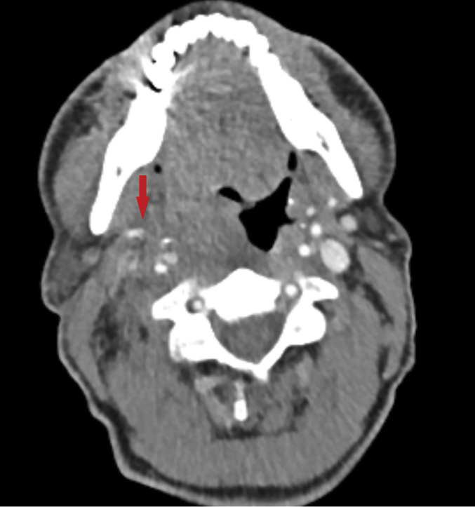

Pre-Operative Computed Tomography Angiography With the Axial Plane Image of the Contrast Blush on the Right Side
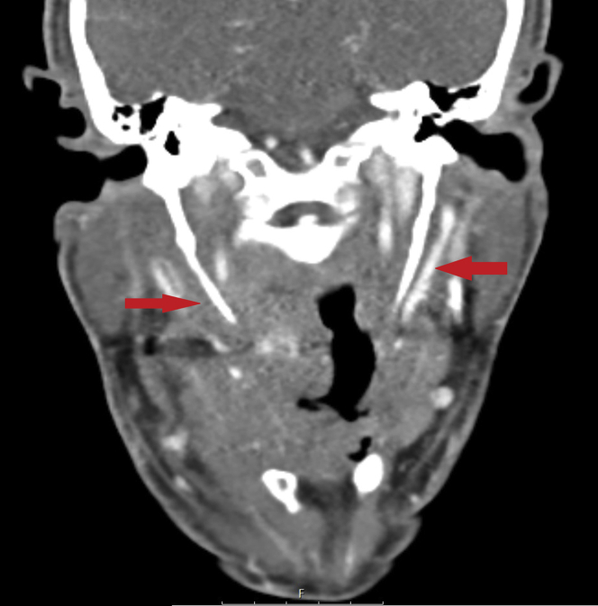
Pre-Operative Computed Tomography Angiography with the Coronal Plane Image of the Bilateral Elongated Styloid Processes
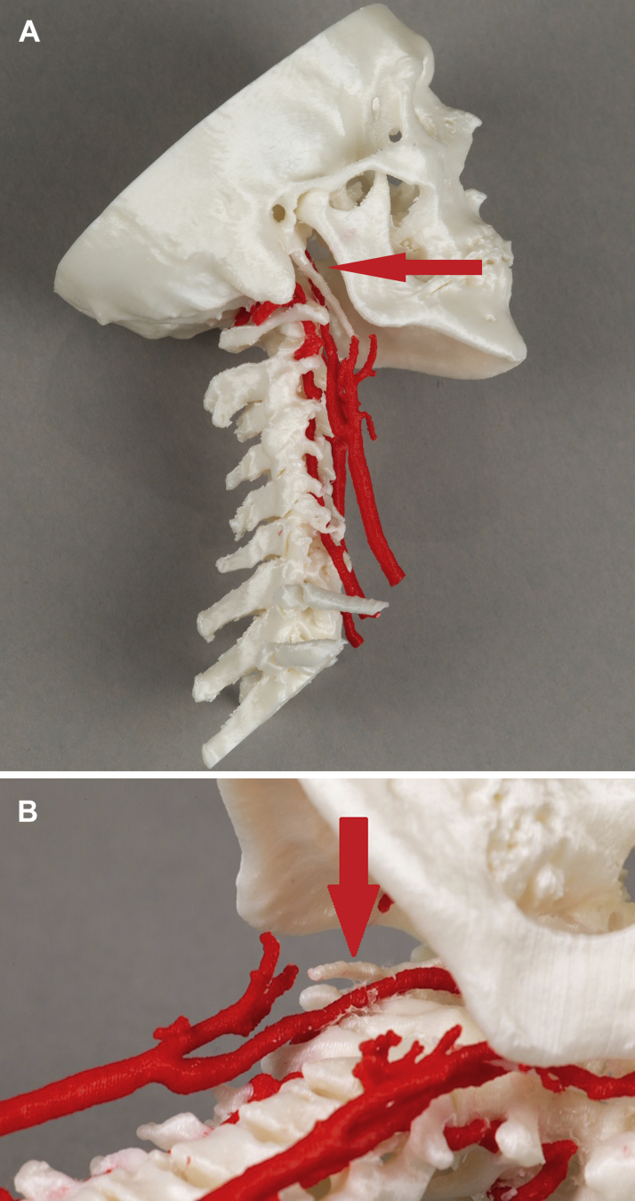
In-House-Printed 3D Model of the Patient's Carotid Arteries and Bony Structures (A) Straight lateral view from the right and (B) an oblique caudocranial view from the left. The in-house-printed 3D model of the patient's carotid arteries and bony structures allows a better understanding of the relationship between the internal carotid artery and styloid process on the right side.
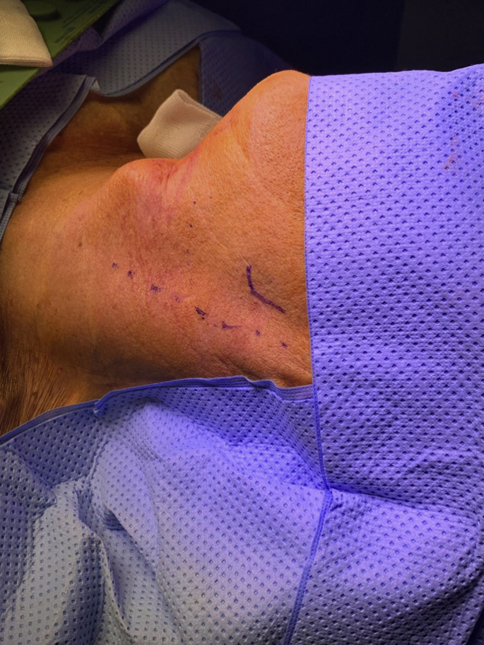
Incision on the Left Side of the Patient’s Mandibular Edge and Ventral Side of the Sternocleidomastoid Muscle Full blue line shows the patient's mandibular edge. The dotted blue line shows the ventral side of the sternocleidomastoid muscle.
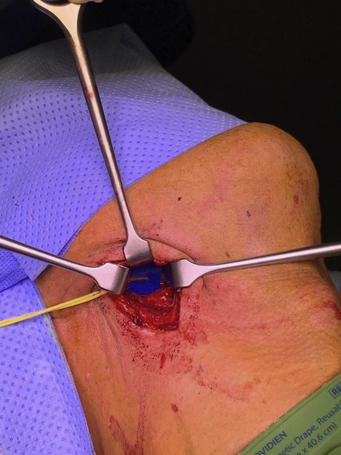
Elongated Styloid Process on the Right Side
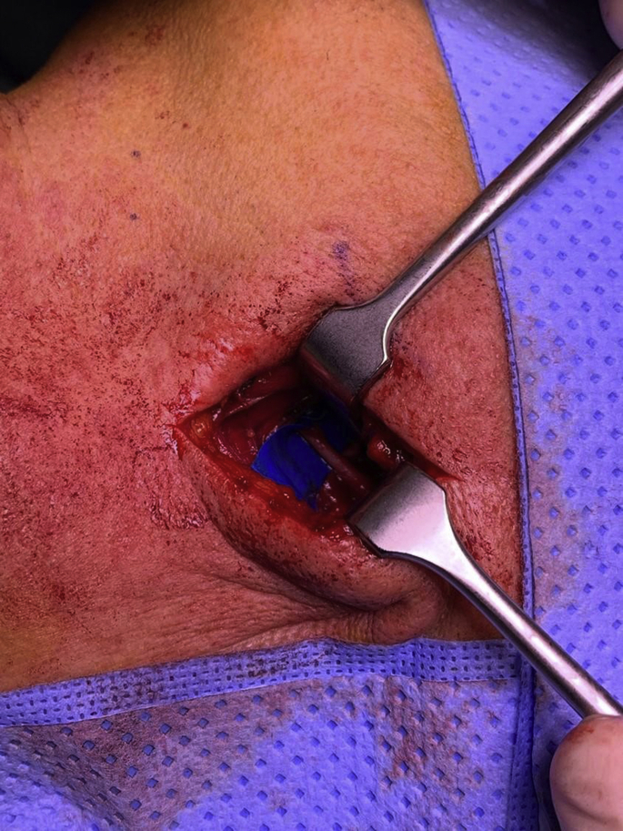
Elongated Styloid Process on the Left Side
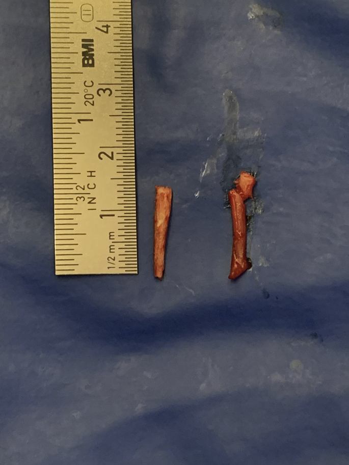
Bilateral Resected Styloid Processes
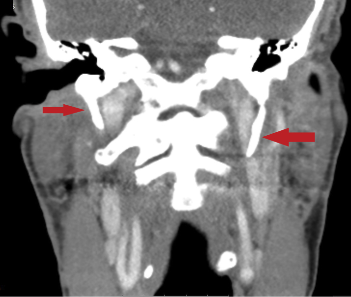
Image in the Coronal Plane from the Post-Operative Computed Tomography Angiography With the Resected Bilateral Styloid Processes The red arrows indicate the location of where the, now resected, styloid proccesi used to be.
References
-
- Eagle W. Elongated styloid processes: Report of Two Cases. Arch Otolaryngol Head Neck Surg. 1937;25:584–587.
-
- Badhey A., Jategaonkar A., Kovacs A., Kadakia S. Eagle syndrome: a comprehensive review. Clin Neurol Neurosurg. 2017;159:34–38. - PubMed
Publication types
LinkOut - more resources
Full Text Sources
