

Case Reports
doi: 10.1016/j.jaccas.2020.03.034.
Massive Myocardial Calcium Deposition: Hardened Heart
Affiliations
- PMID: 34317401
- PMCID: PMC8302108
- DOI: 10.1016/j.jaccas.2020.03.034
Item in Clipboard
Case Reports
Massive Myocardial Calcium Deposition: Hardened Heart
JACC Case Rep.
.
Abstract
A 25-year-old African-American woman with end-stage renal disease presented with new-onset heart failure. Transthoracic echocardiography indicated a significantly hyperechoic myocardium, and computed tomography noted a circumferential hyperattenuated myocardium. Endomyocardial biopsy revealed focal interstitial and intramyocyte calcium deposition in the heart, confirming a rare diagnosis of massive myocardial calcium deposition. (Level of Difficulty: Beginner.).
Keywords: CT, computed tomographic; EMB, endomyocardial biopsy; ESRD, end-stage renal disease; HF, heart failure; cardiomyopathy; disorders of calcium metabolism; restrictive.
© 2020 The Authors.
Figures


Electrocardiogram

Echocardiogram (A) Pulsed-wave Doppler through the mitral valve. (B) Tissue Doppler of the medial mitral valve annulus. (C) Tissue Doppler of the lateral mitral valve annulus. DT = E-wave deceleration time; E = E wave; e′ = e′ mitral velocity.

Computed Tomography Imaging of the Chest (A) Computed tomographic (CT) imaging of the chest. Arrows denote calcified myocardium. (B) Contrast-enhanced CT imaging of the chest with multiplanar reformatting in the 4-chamber view. (C) Contrast-enhanced CT imaging of the chest with multiplanar reformatting in the short-axis view. A = calcified myocardium; B = noncalcified endocardium; LA = left atrium; LV = left ventricle; RA = right atrium; RV = right ventricle.
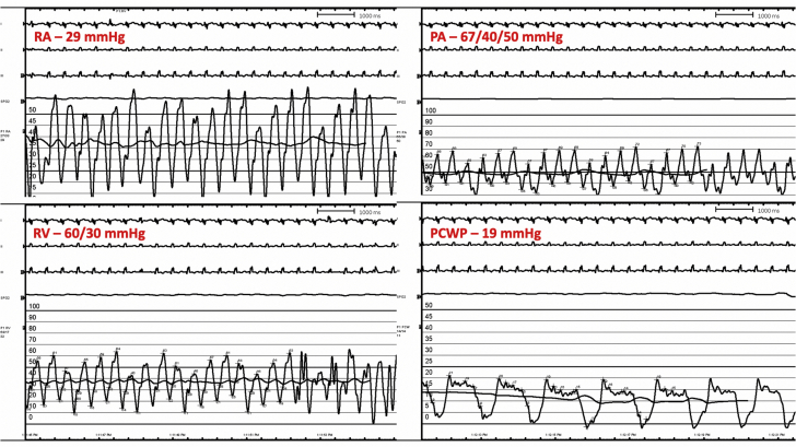
Right Heart Catheterization Tracings PA = pulmonary artery pressure; PCWP = pulmonary capillary wedge pressure; RA = right atrial pressure; RV = right ventricular pressure.
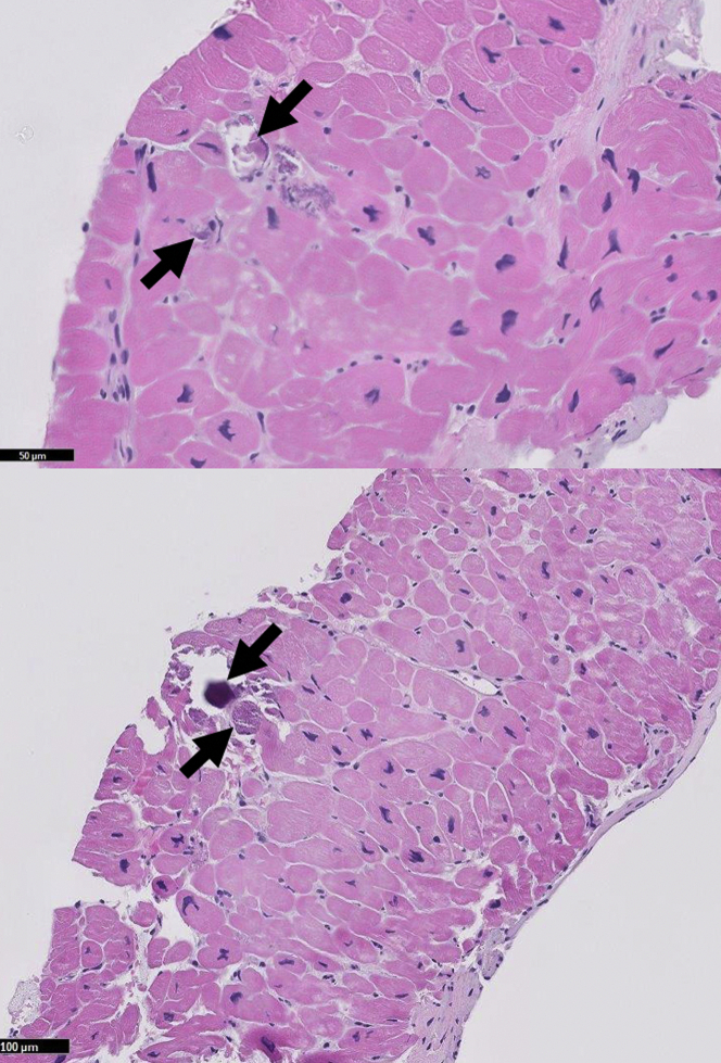
Endomyocardial Biopsy With Routine Stain Arrows denote intracellular calcium deposits.

Endomyocardial Biopsy With Von Kossa Stain Arrows denote intracellular calcium deposits.

Pathophysiology of Secondary Hyperparathyroidism CKD = chronic kidney disease; GI = gastrointestinal; PTH = parathyroid hormone.
References
-
- Na J.Y. A heart of stone: an autopsy case of massive myocardial calcification. Forensic Sci Med Pathol. 2018;14:102–105. - PubMed
-
- Isotalo P.A., Halil A., Green M., Tang A., Lach B., Veinot J.P. Metastatic calcification of the cardiac conduction system with heart block: an under-reported entity in chronic renal failure patients. J Forensic Sci. 2000;45:1335–1338. - PubMed
Publication types
Grants and funding
LinkOut - more resources
Full Text Sources
Research Materials
Miscellaneous
