

Case Reports
doi: 10.1016/j.jaccas.2020.05.076.
eCollection 2020 Jul.
Repair of Spontaneous Left Atrial Dissection Resulting in Severe Paravalvular Native Mitral Valve Regurgitation
Affiliations
- PMID: 34317424
- PMCID: PMC8311892
- DOI: 10.1016/j.jaccas.2020.05.076
Item in Clipboard
Case Reports
Repair of Spontaneous Left Atrial Dissection Resulting in Severe Paravalvular Native Mitral Valve Regurgitation
JACC Case Rep.
.
Abstract
A 54-year-old male with history of end-stage renal disease secondary to hypertension on hemodialysis with moderate aortic valve insufficiency presented with progressive exertional dyspnea and lower extremity edema over several weeks. Relevant history included hospitalization for Staphylococcus epidermidis bacteremia secondary to dialysis catheter line infection 6 months prior. (Level of Difficulty: Advanced.).
Keywords: aortic valve; endocarditis; mitral valve.
© 2020 The Authors.
Figures

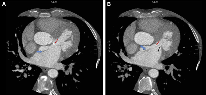
Coronary Computed Tomography Angiogram Demonstrating the Left Atrial Dissection (A) Single-axial image from a coronary computed tomography angiogram (CTA) showing calcification of the aorticomitral curtain (red arrow) that is secondary to a chronic aortic insufficiency jet. This lesion served as the nidus for the dissection (blue arrow). (B) Single-axial image from a coronary CTA showing the left atrial dissection flap (blue arrow) and native perivalvular mitral regurgitation due to anterior mitral leaflet (black arrow) dehiscence.
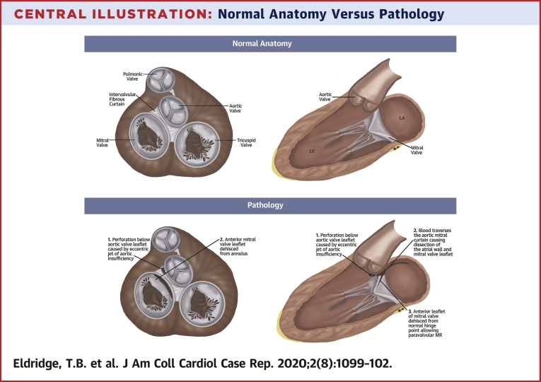
Normal Anatomy Versus Pathology Disruption of the aorto-mitral continuity causing a large dissection plane with disruption of the adventitia at the base of the aorta near the aortic valve annulus extending approximately 6 cm along the left atrial wall. Systolic flow was noted within this space with an exit point approximately 5 cm from the origin and flow continuing into the left atrium (LA) resulting in severe paravalvular native valve mitral regurgitation. The perforation below the aortic leaflet (1) induced blood flow across the aorto-mitral curtain causing dissection of the atrial wall and mitral leaflet (2). Subsequently, the anterior mitral leaflet dehisced allowing significant paravalvular leak (3). LV = left ventricle.
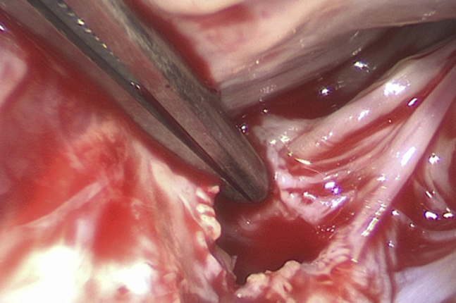
A Subannular Perforation of the Aorticomitral Curtain at the Base of the Anterior Mitral Leaflet
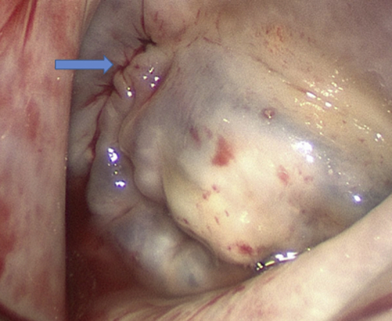
The Dome of the Left Atrium The arrow indicates the aorticomitral curtain left atrial dissection flap.
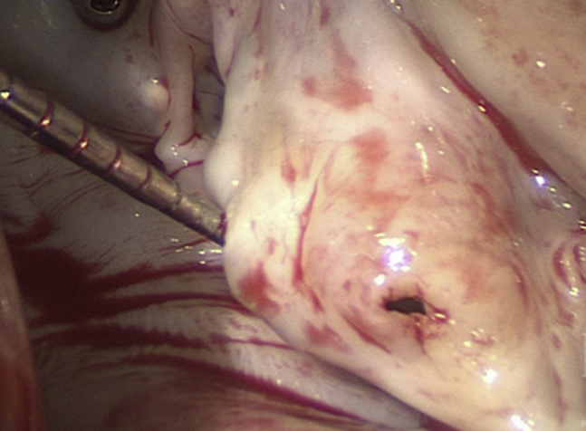
Perforation of the Anterior Mitral Leaflet
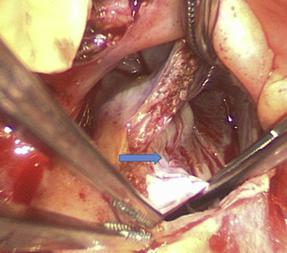
Mitral Valve Saline Test After Leaflet Repair The arrow indicates the suture line from the aorticomitral curtain patch.
References
Publication types
LinkOut - more resources
Full Text Sources
