

Case Reports
doi: 10.1016/j.jaccas.2020.11.038.
eCollection 2021 Jan.
An Unusual Complication of Transseptal Puncture
Affiliations
- PMID: 34317466
- PMCID: PMC8305646
- DOI: 10.1016/j.jaccas.2020.11.038
Item in Clipboard
Case Reports
An Unusual Complication of Transseptal Puncture
JACC Case Rep.
.
Abstract
The interatrial septum is a structure with complex embryological development. The true atrial septum is a circumscribed structure, and transgression outside of this area during transseptal puncture may result in entry into the extracardiac space or aorta that may result in a pericardial effusion or cardiac tamponade. (Level of Difficulty: Intermediate.).
Keywords: IAS, interatrial septum; SVC, superior vena cava; atrial fibrillation; complication; electrophysiology; pericardial effusion.
© 2021 The Authors.
Conflict of interest statement
The authors have reported that they have no relationships relevant to the contents of this paper to disclose.
Figures

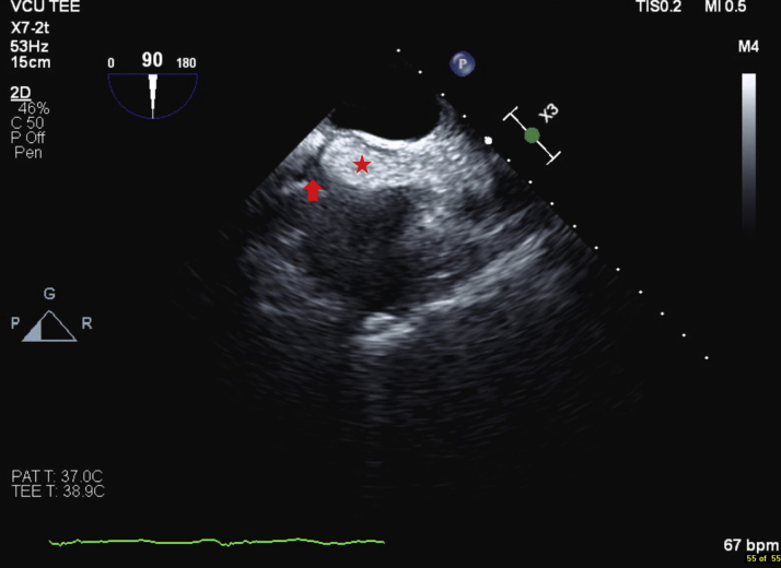
Transesophageal Echocardiography Bicaval View (Star) Interatrial septum hypertrophy. Sparing of the fossa ovalis (arrow).
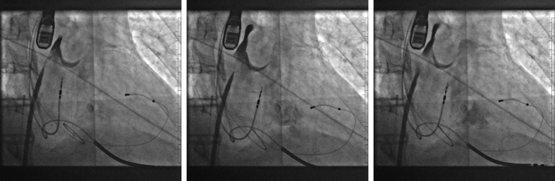
Right and Left Anterior Oblique Projections of Wire Advancement Following Transseptal Puncture

Left Atrial Pressure Recording
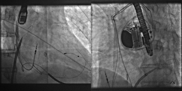
Right Anterior Oblique Projection of Contrast Extravasation Into Transverse Pericardial Space and Simultaneous Contrast Flow Into the Left Ventricle
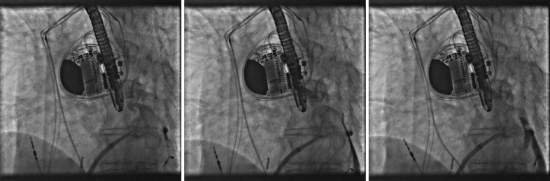
Left Anterior Oblique Projection of Contrast Extravasation Into Transverse Pericardial Space
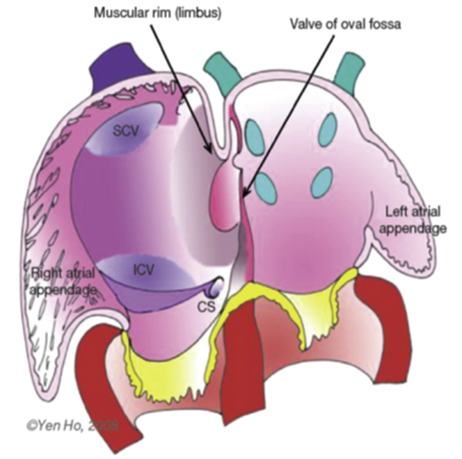
Component Parts of the Definitive Atrial Septum IVC = inferior vena cava; SVC = superior vena cava.
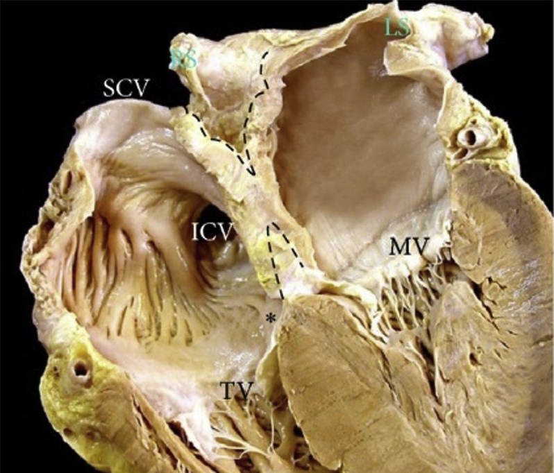
4-Chamber Section Through the Heart Four-chamber section through the heart showing the offset arrangement of the mitral valve (MV) and tricuspid valve (TV) which produces the so-called muscular atrioventricular septum (∗) and the deep in folding (dotted lines) of the atrial wall superior and inferior to the floor of the oval fossa. IVC = inferior vena cava; SVC = superior vena cava.
Similar articles
-
Transseptal Puncture Through an Interatrial Septum With Lipomatous Hypertrophy: A False Perception of Success and Failure.Cureus. 2022 Sep 29;14(9):e29737. doi: 10.7759/cureus.29737. eCollection 2022 Sep. Cureus. 2022. PMID: 36324361 Free PMC article.
-
Interatrial septum dissection and atrial wall hematoma following transseptal puncture: A systematic review of the literature.Catheter Cardiovasc Interv. 2020 Aug;96(2):424-431. doi: 10.1002/ccd.28554. Epub 2019 Oct 23. Catheter Cardiovasc Interv. 2020. PMID: 31642609
-
Transseptal access for left atrial ablation: the catheter-probing techniques are not without risk.J Cardiovasc Electrophysiol. 2014 May;25(5):479-484. doi: 10.1111/jce.12356. Epub 2014 Jan 29. J Cardiovasc Electrophysiol. 2014. PMID: 24384060
-
Transseptal puncture and catheter ablation via the superior vena cava approach for persistent atrial fibrillation in a patient with polysplenia syndrome and interruption of the inferior vena cava: contact force-guided pulmonary vein isolation.Europace. 2017 Jul 1;19(7):1227-1232. doi: 10.1093/europace/euw095. Europace. 2017. PMID: 27174901
-
Current Concepts of Anatomy, Electrophysiology, and Therapeutic Implications of the Interatrial Septum.JACC Clin Electrophysiol. 2019 Jun;5(6):647-656. doi: 10.1016/j.jacep.2019.04.013. JACC Clin Electrophysiol. 2019. PMID: 31221350 Review.
Cited by
-
Inadvertent left atrial injury during transseptal puncture with a radiofrequency-powered wire.J Interv Card Electrophysiol. 2023 Oct;66(7):1561-1562. doi: 10.1007/s10840-023-01560-6. Epub 2023 May 27. J Interv Card Electrophysiol. 2023. PMID: 37237134 No abstract available.
-
Interatrial septum dissection and closure from transseptal puncture during mitral transcatheter edge-to-edge repair: a case report.Eur Heart J Case Rep. 2024 Nov 2;8(11):ytae559. doi: 10.1093/ehjcr/ytae559. eCollection 2024 Nov. Eur Heart J Case Rep. 2024. PMID: 39545161 Free PMC article.
-
Delayed Tamponade After Transseptal Puncture: Focus on the Mechanism of a Rare But Life-Threatening Complication.JACC Case Rep. 2025 Apr 2;30(7):103379. doi: 10.1016/j.jaccas.2025.103379. JACC Case Rep. 2025. PMID: 40185610 Free PMC article.
References
-
- January C.T., Wann L.S., Calkins H. 2019 AHA/ACC/HRS focused update of the 2014 AHA/ACC/HRS guideline for the management of patients with atrial fibrillation: a report of the American College of Cardiology/American Heart Association Task Force on Clinical Practice Guidelines and the Heart Rhythm Society. J Am Coll Cardiol. 2019;74:104–132. - PubMed
-
- Cournand A.M.H. Recording of blood pressure from the left auricle and the pulmonary veins in human subjects with interauricular septal defect. Am J Physiol. 1947;150:267–271. - PubMed
-
- Brockenbrough E.C., Braunwald E.R.J. Transseptal left heart catheterization. A review of 450 studies and description of an improved technic. Circulation. 1962;25:15–21. - PubMed
-
- Katritsis G.D., Siontis G.C.M., Giazitzoglou E., Fragakis N., Katritsis D.G. Complications of transseptal catheterization for different cardiac procedures. Int J Cardiol. 2013;168:5352–5354. - PubMed
-
- Anderson R.B.N. The anatomy of the heart revisited. Anat Rec. 1996;246:1–7. - PubMed
Publication types
LinkOut - more resources
Full Text Sources
