Case Reports
. 2020 Dec 9;3(1):104-108.
doi: 10.1016/j.jaccas.2020.09.039. eCollection 2021 Jan.

The Spectrum of Caseous Mitral Annulus Calcifications

Affiliations
Case Reports

The Spectrum of Caseous Mitral Annulus Calcifications

Agnes Mayr et al. JACC Case Rep. .

Abstract

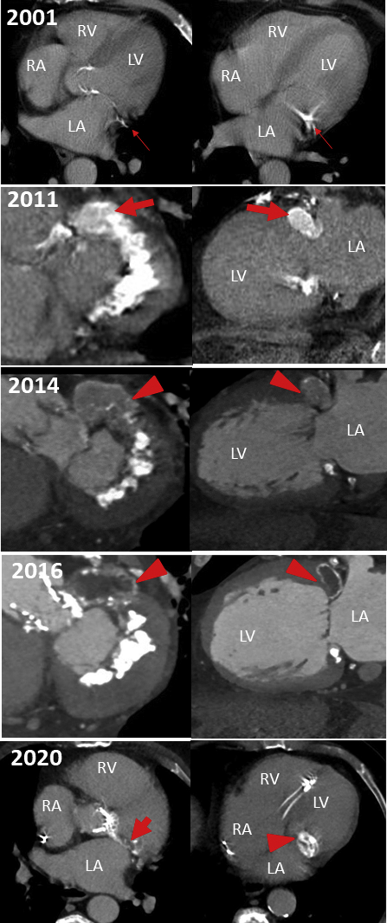
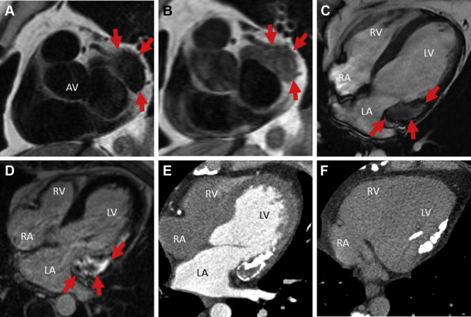
Mitral annular calcification (MAC) is a chronic, degenerative condition of the fibrous mitral annulus, which may transform to liquefaction necrosis MAC, a rare variant of caseous MAC. We present a series of experiences, showing the varying manifestations of caseous MAC according to multimodal imaging. (Level of Difficulty: Intermediate.).

Keywords: CMAC, caseous mitral annular calcification; CMR, cardiac magnetic resonance; CT, computed tomography; MAC, mitral annular calcification; TAVI, transcatheter aortic valve intervention; cardiac magnetic resonance imaging; caseous mitral annular calcification; computed tomography; liquefaction necrosis.


Conflict of interest statement

The authors have reported that they have no relationships relevant to the contents of this paper to disclose.

Figures

None
Graphical abstract
Figure 1
Figure 1
Dynamic Evolution and Atypical Location of a Caseous Mitral Annular Calcification LA = left atrium; LV = left ventricle; RA = right atrium; RV = right ventricle.
Figure 2
Figure 2
Atypical Mobile Caseous Mitral Annular Calcification Mimicking a Valvular Mass (A) Apical 4-chamber view of transesophageal echocardiography. (B) Apical 4-chamber tissue Doppler in transesophageal echocardiography. (C) Apical 4-chamber view of transthoracic 3D echocardiography. (D) Biphasic iodine contrast-enhanced electrocardiographically gated computed tomography (CT). (E) Basal short-axis slice of late gadolinium enhancement cardiac magnetic resonance imaging. AV = aortic valve; other abbreviations as in Figure 1.
Figure 3
Figure 3
Caseous Mitral Annular Calcification at Cardiac Magnetic Resonance Imaging (A) Basal short-axis slice of a T2-weighted turbo-spin-echo cardiac magnetic resonance imaging sequence. (B) Basal short-axis slice of a T1-weighted turbo-spin-echo cardiac magnetic resonance imaging sequence. (C) 4-chamber view of a cine steady-state free precession cardiac magnetic resonance imaging sequence. (D) 4-chamber view of a late gadolinium enhancement cardiac magnetic resonance imaging sequence. (E) Iodine contrast-enhanced electrocardiographically gated computed tomography (CT). (F) Contrast-free electrocardiographically gated CT. Abbreviations as in Figures 1 and 2.

References

    1. Deluca G., Correale M., Ieva R., Del Salvatore B., Gramenzi S., Di Biase M. The incidence and clinical course of caseous calcification of the mitral annulus: a prospective echocardiographic study. J Am Soc Echocardiogr. 2008;21:828–833. - PubMed
    1. Pomerance A. Pathological and clinical study of calcification of the mitral valve ring. J Clin Pathol. 1970;23:354–361. - PMC - PubMed
    1. Motwani M., Fairbairn T.A., Jogiya R., Greenwood J.P., Plein S. Caseous calcification of the mitral valve complicated by embolization, mitral regurgitation, and pericardial constriction. Eur Heart J Cardiovasc Imaging. 2012;13:792. - PubMed
    1. Asselbergs F.W., Mozaffarian D., Katz R. Association of renal function with cardiac calcifications in older adults: the Cardiovascular Health Study. Nephrol Dial Transplant. 2009;24:834–840. - PMC - PubMed
    1. Koito H., Nakamura C., Suzuki J., Takahashi H., Iwasaka T. Reduced size of liquefaction necrosis of mitral annular calcification in chronic renal failure by using low calcium concentration hemodialysis. Jpn Circ J. 1999;63:490–492. - PubMed

Publication types

LinkOut - more resources