

Fulminant Eosinophilic Myocarditis and VT Storm
- PMID: 34317561
- PMCID: PMC8311013
- DOI: 10.1016/j.jaccas.2020.12.028
Fulminant Eosinophilic Myocarditis and VT Storm
Abstract
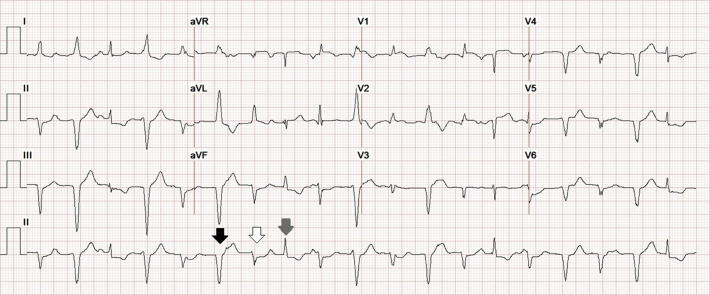
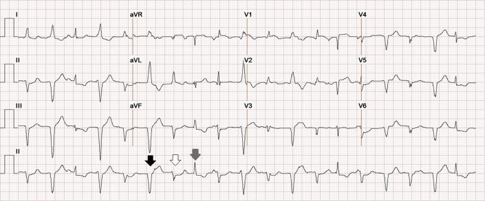
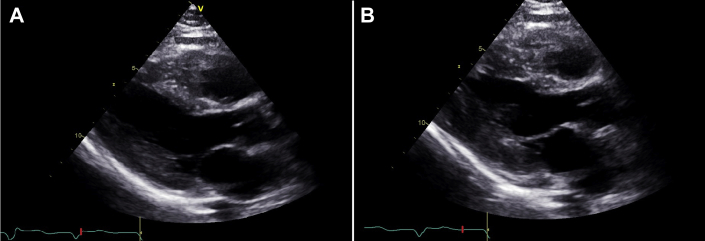
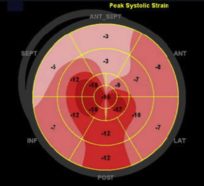
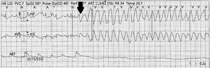
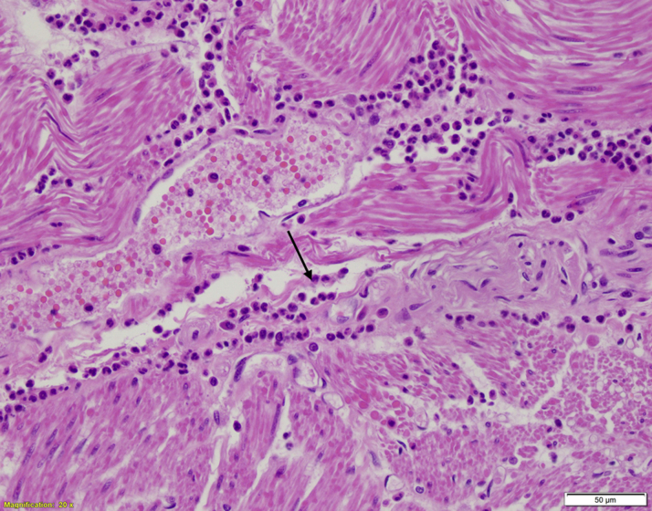
Eosinophilic myocarditis is a rare and frequently fatal disease that is often undiagnosed until autopsy. We report a case of eosinophilic myocarditis with an unusual initial presentation of palpitations that subsequently evolved into ventricular tachycardia storm and death within 4 days. (Level of Difficulty: Beginner.).
Keywords: EM, eosinophilic myocarditis; PVC, pre-mature ventricular contractions; VF, ventricular fibrillation; VT, ventricular tachycardia; eosinophilic myocarditis; palpitations; polymorphic pre-mature ventricular contractions; ventricular tachycardia storm.
© 2021 The Authors.
Conflict of interest statement
The authors have reported that they have no relationships relevant to the contents of this paper to disclose.
Figures
References
-
- Roberts W.C., Kietzman A.T. Severe eosinophilic myocarditis in the portion of the left ventricular wall excised to insert a left ventricular assist device for severe heart failure. Am J Cardiol. 2020;125:264–269. - PubMed
-
- Brambatti M., Matassini M.V., Adler E.D. Eosinophilic myocarditis: characteristics, treatment, and outcomes. J Am Coll Cardiol. 2017;70:2363–2375. - PubMed
-
- Chaudhry M.A., Grazette L., Yoon A. Churg-Strauss syndrome presenting as acute necrotizing eosinophilic myocarditis: concise review of the literature. Curr Hypertens Rev. 2014;15:8–12. - PubMed
-
- Kociol R.D., Cooper L.T., Fang J.C. Recognition and initial management of fulminant myocarditis: a scientific statement from the American Heart Association. Circulation. 2020;141:e69–e92. - PubMed
Publication types
LinkOut - more resources
Full Text Sources
