

Acute Mitral Regurgitation Secondary to Spontaneous Left Atrial Appendage Occluder Migration
- PMID: 34317648
- PMCID: PMC8311284
- DOI: 10.1016/j.jaccas.2021.01.016
Acute Mitral Regurgitation Secondary to Spontaneous Left Atrial Appendage Occluder Migration
Abstract
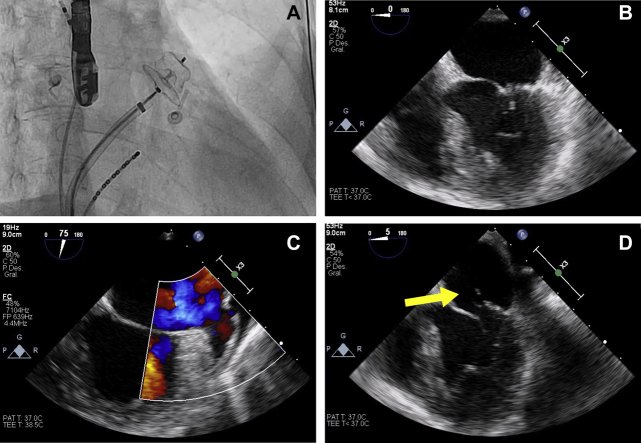
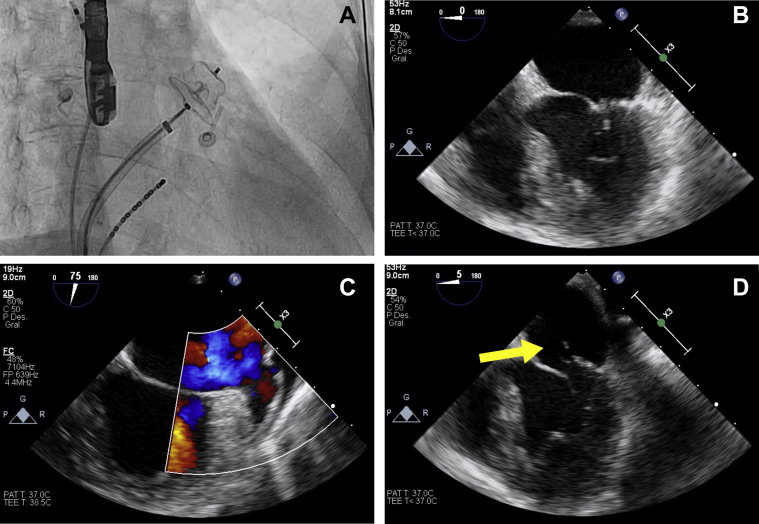
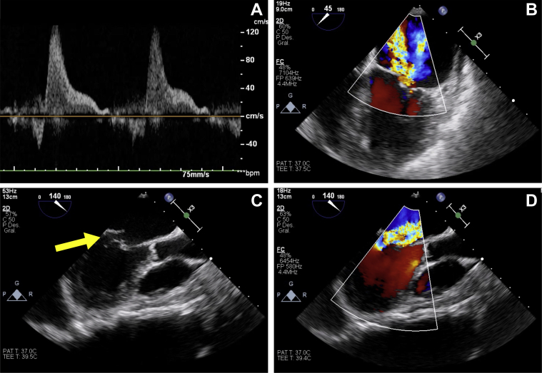
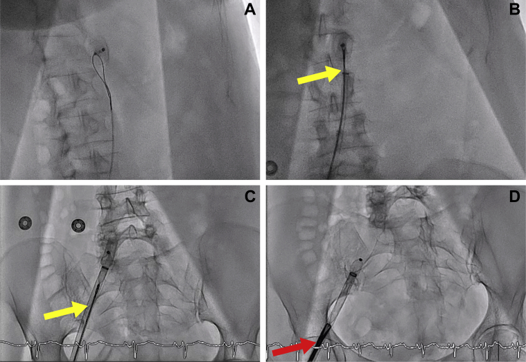
A patient underwent left atrial appendage occlusion due to recurrent stroke despite new oral anticoagulant therapy. The patient later presented with severe acute mitral regurgitation secondary to occluder device migration, which was retrieved percutaneously from the descending aorta via the femoral artery. Mitral surgical repair was required and successfully performed. (Level of Difficulty: Intermediate.).
Keywords: AF, atrial fibrillation; LAAO, left atrial appendage occlusion; OAC, oral anticoagulant; TOE, transesophageal echocardiography; acute heart failure; atrial fibrillation; mitral valve; stroke.
© 2021 The Authors.
Conflict of interest statement
The authors have reported that they have no relationships relevant to the contents of this paper to disclose.
Figures
References
-
- Pison L., Potpara T.S., Chen J. Left atrial appendage closure-indications, techniques, and outcomes: results of the European Heart Rhythm Association Survey. Europace. 2015;17:642–666. - PubMed
-
- Thambidorai S.K., Murray R.D., Parakh K. Utility of transesophageal echocardiography in identification of thrombogenic milieu in patients with atrial fibrillation (an ACUTE ancillary study) Am J Cardiol. 2005;96:935–941. - PubMed
-
- Cruz-González I., González-Ferreiro R., Freixa X. Left atrial appendage occlusion for stroke despite oral anticoagulation (resistant stroke). Results from the Amplatzer Cardiac Plug registry. Rev Esp Cardiol (Engl Ed) 2020;73:28–34. - PubMed
Publication types
LinkOut - more resources
Full Text Sources
