

Muscle-sparing aortic coarctation repair
- PMID: 34317891
- PMCID: PMC8302918
- DOI: 10.1016/j.xjtc.2020.05.005
Muscle-sparing aortic coarctation repair
Abstract
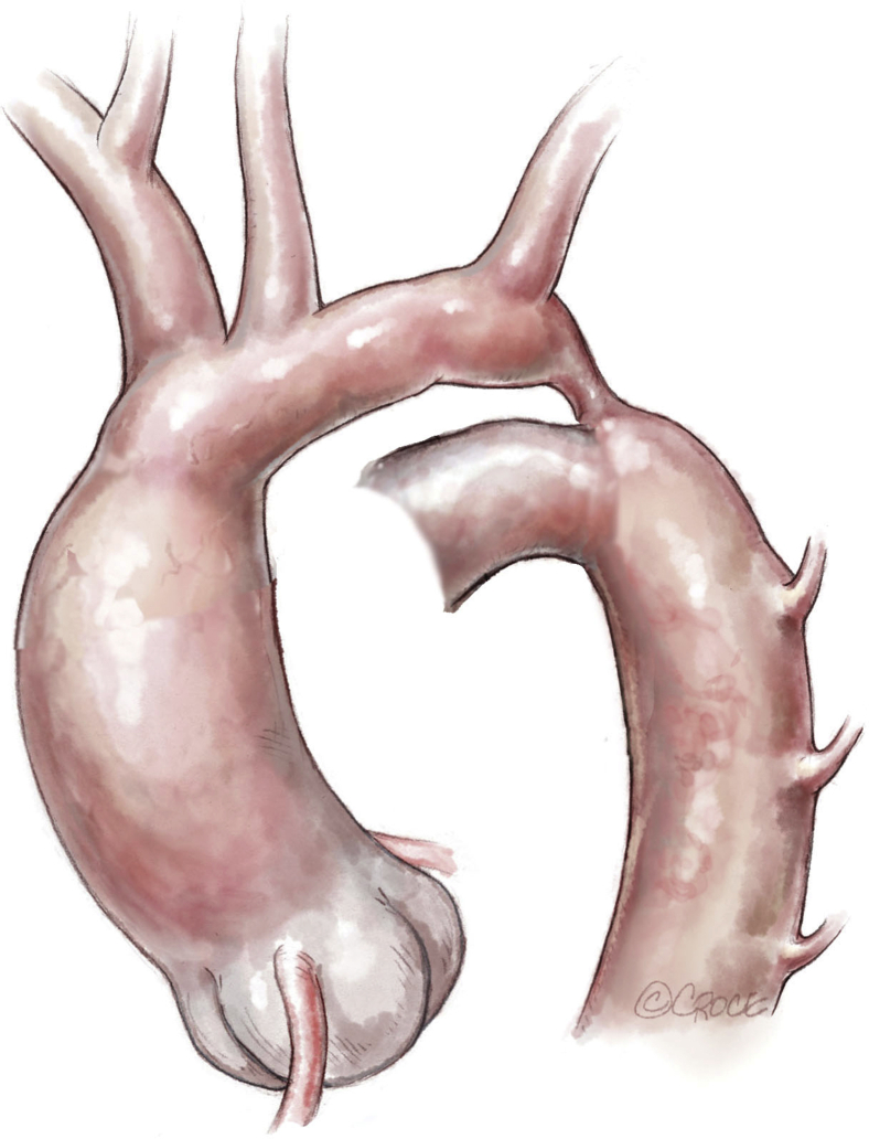
Objective: Surgery for aortic coarctation repair provides excellent hemodynamic results but may be complicated by musculoskeletal issues. The purpose of the study was to determine the midterm results of a muscle-sparing surgical approach to aortic coarctation repair, with special emphasis on the repair and on the musculoskeletal changes associated with a posterior thoracotomy.
Methods: We included all children with aortic coarctation operated on with our minimally invasive approach between June 2002 and October 2004, with a follow-up of ≥4.5 years. Patients were assessed clinically and echocardiographically. The spine, left chest, and shoulder were assessed clinically and radiographically.
Results: Thirty-one children were included. The age at operation ranged from 1 day to 15 months and weight ranged from 980 g to 10 kg. All patients underwent an extended end-to-end anastomosis coarctation repair through a minimal (n = 19) or total-muscle sparing (n = 12) or extrapleural (n = 18) approach. Five patients had an additional enlargement procedure on the aortic arch. 27 patients had no residual or recurrent gradient. Four patients exhibited restenosis, for which 1 underwent a percutaneous angioplasty and 2 underwent surgical reintervention. All patients were free of hypertension. One patient had borderline values. The musculoskeletal assessment was normal in all but 3 patients. Two patients who underwent other subsequent thoracic surgeries developed thoracogenic scoliosis of moderate severity. A third patient had a left winged scapula. No rib fusion or intercostal space enlargement was found.
Conclusions: Compared with a conventional approach, our minimally invasive surgical approach led to excellent musculoskeletal outcomes without compromising the hemodynamic results.
Keywords: children; coarctation of the aorta; muscle-sparing approach.
© 2020 The Authors.
Figures


References
-
- Dave H.H., Buechel E.R.V., Prêtre R. Muscle-sparing extrapleural approach for the repair of aortic coarctation. Ann Thorac Surg. 2006;81:243–248. - PubMed
-
- Bal S., Elshershari H., Celiker R., Celiker A. Thoracic sequels after thoracotomies in children with congenital cardiac disease. Cardiol Young. 2003;13:264–267. - PubMed
-
- Emmel M., Ulbach P., Herse B., Dalichau H., Haupt W.F., Schumann D., et al. Neurogenic lesions after posterolateral thoracotomy in young children. Thorac Cardiovasc Surg. 1996;44:86–91. - PubMed
-
- Roclawski M., Pankowski R., Smoczynski A., Ceynowa M., Kloc W., Wasilewski W., et al. Secondary scoliosis after thoracotomy in patients with aortic coarctation and patent ductus arteriosus. Stud Health Technol Inform. 2012;176:43–46. - PubMed
-
- Van Biezen F.C., Bakx P.A., De Villeneuve V.H., Hop W.C. Scoliosis in children after thoracotomy for aortic coarctation. J Bone Jt Surg Am. 1993;75:514–518. - PubMed
LinkOut - more resources
Full Text Sources
