. 2020 Aug 1:4:208-216.
doi: 10.1016/j.xjtc.2020.07.027. eCollection 2020 Dec.

Scimitar syndrome: A new multipatch technique and incidence of postoperative pulmonary vein obstruction

Affiliations

Scimitar syndrome: A new multipatch technique and incidence of postoperative pulmonary vein obstruction

Robert L Geggel et al. JTCVS Tech. .

Abstract

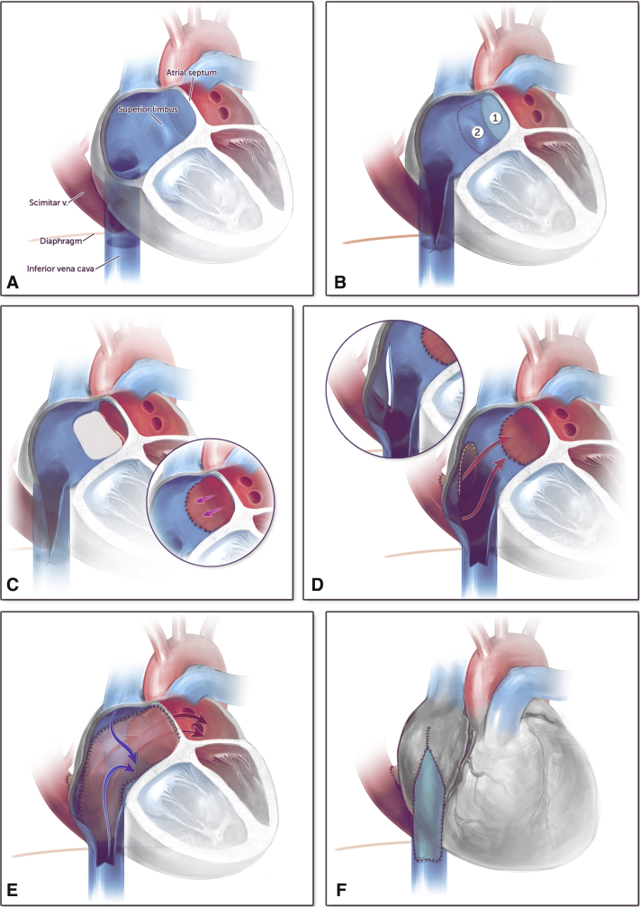
Objective: A review of our center's experience before March 2011 showed that one half of 36 patients who had a baffling or reimplantation procedure to repair scimitar syndrome developed pulmonary vein obstruction. We analyzed the results of a new operation that enlarges the left atrium and avoids circuitous pathways or tension on the scimitar pulmonary vein.

Methods: Between April 2011 and November 2018, 22 patients underwent scimitar vein surgery; 11 had baffling or reimplantation and 11 only had the new operation that included resection of the atrial septum with removal of the muscular limbus. The left atrium was pulled down toward the scimitar vein and a V-shaped incision made at the scimitar vein atrial junction with the space filled with a pulmonary homograft. If the scimitar vein coursed adjacent to the atrium, a V-shaped incision was made into the scimitar vein and directly anastomosed to the atrium. A patch of autologous pericardium was used to septate the atrium and an additional patch placed anteriorly to augment the inferior vena cava.

Results: Of the 11 patients who had baffling or reimplantation, 5 developed pulmonary vein obstruction between 45 days and 9.5 months after surgery associated with baffle thrombosis or tension on the pulmonary vein. None of the 11 patients who only had the new procedure developed pulmonary vein obstruction during postoperative monitoring up to 3.6 years.

Conclusions: Patients having only the multipatch procedure for repair of scimitar syndrome have not developed postoperative pulmonary vein obstruction in the short to intermediate term.

Keywords: CMR, cardiac magnetic resonance; IVC, inferior vena cava; congenital heart disease; partial anomalous pulmonary venous return; pulmonary vein stenosis; scimitar syndrome.


Figures

None
New surgical technique enlarges left atrium and avoids kinking or tension on pathway.
Figure 1
Figure 1
Sites of obstruction associated with traditional baffle repair of scimitar syndrome. Diagram depicts traditional baffle repair of scimitar syndrome. The stars depict sites of potential obstruction associated with the long angulated course of the scimitar vein baffle pathway, including at the proximal segment of the scimitar vein baffle pathway, superior limbic band, and atrial septal defect.
Figure 2
Figure 2
Triple patch operative technique to repair scimitar syndrome. A, Baseline anatomy showing scimitar vein connecting to the IVC superior to the level of the diaphragm and intact atrial septum. B, An incision is made in the right atrium and IVC to identify the origin of the scimitar vein. The atrial septum (1) and superior limbic band (2) are imaged. C, The atrial septum and superior limbic band have been resected. The left atrium is pulled downward and anastomosed to the right atrial tissue bringing the left atrium closer to the scimitar vein. D, A V-shaped incision is made in the atrium and scimitar vein and the space is filled in with a thin pulmonary homograft. E, An autologous pericardial patch is placed to divide the scimitar vein from the IVC and direct flow from the scimitar vein via a relatively straight baffle to the left atrium (red arrows) effectively closing the atrial septal defect. The vena cava flow courses anterior to this patch (blue arrows). F, A pericardial patch is placed in the IVC in the region where the baffle patch was placed to “raise the roof” and augment the caliber of this region.
Figure 3
Figure 3
Preoperative cineangiograms. Preoperative anteroposterior and lateral views of levophase of right pulmonary artery angiogram in a 7-year-old patient who had the SV coursing a distance from the atrium and who subsequently had the triple multipatch procedure using pulmonary homograft insertion to connect the SV to the atrial tissue (A, B) and in a 10-year-old patient who had the SV in close proximity to the atrium and who subsequently had direct connection of the SV to the atrial tissue without use of pulmonary homograft material (C, D). SV, Scimitar vein.
Video 1
Video 1
Before establishing cardiopulmonary bypass, the scimitar vein and inferior vena cava are widely mobilized off the pericardial reflection laterally and inferiorly along the diaphragm. Cardiopulmonary bypass is established and the right atrium is opened. The right atriotomy is extended superiorly and then inferiorly. The intracardiac anatomy is explored demonstrating the scimitar vein laterally. The interatrial septum is incised entering the left atrium. The atrial septal tissue is then extensively resected including the superior limbus and the muscular portion of the interatrial septum. This resection includes the back wall of the left atrium thus exiting the heart posteriorly. The back wall of the left atrium is brought rightward and laterally toward the scimitar vein and sewn in place with a running 6-0 polypropylene suture, which effectively shortens the distance between the scimitar vein and the left atrium. The lateral portion of the back wall of the right atrium is incised. This incision is extended inferiorly into the scimitar vein. A patch of pulmonary homograft is fashioned and sewn into the back wall of the left atrium and pulmonary venous pathway to fill in the gap created by the incision. A patch of autologous pericardium is cut to be used to close the atrial septal defect and baffle the scimitar vein to the left atrium. The suture line starts at the junction of the inferior vena cava and the scimitar vein and is carried superiorly and laterally along the ridge of the scimitar vein. The patch is large enough so there is a patulous pathway between the scimitar vein and left atrium. The superior aspect of the patch is closed. The inferior vena cava cannula is then removed and pump suckers are placed in the inferior vena cava to control systemic venous return. The inferior vena cava is incised inferiorly and a patch of autologous pericardium is placed to augment the inferior vena cava to right atrium pathway. The patch is carried up laterally and superiorly along the right atrial wall. After the patch is sewn in place, the right atriotomy is closed primarily with 2 layers of running 5-0 polypropylene suture. Video available at: https://www.jtcvs.org/article/S2666-2507(20)30365-5/fulltext.
Figure 4
Figure 4
Postoperative cardiac computed tomography scan. Postoperative computed tomography scan of 7-year-old child who had the triple multipatch procedure as part of surgical repair. Unobstructed left upper (LU) and left lower (LL) pulmonary veins connecting to the left atrium are shown in (A) and unobstructed scimitar vein (SV) is shown in (B) This patient also had the right upper (RU) pulmonary vein connecting to the azygos vein and had this vein baffled separately to the left atrium. Ao, Aorta.
Figure 5
Figure 5
Freedom from scimitar vein obstruction after surgical repair. Kaplan–Meier curve depicting freedom from development of scimitar vein obstruction after standard repair (baffling or reimplantation) and the new procedure using double or triple patches as described in the text. The variable length of follow-up is depicted by the number of patients at risk over the study period. The shaded area depicts the confidence band for the baffle or reimplantation group. Confidence bands could not be constructed for the new procedure group, since no patients developed obstruction.

References

    1. Neill C.A., Ferencz C., Sabiston D.C., Sheldon H. The familial occurrence of hypoplastic right lung with systemic arterial supply and venous drainage “scimitar syndrome.”. Bull Johns Hopkins Hosp. 1960;107:1–15. - PubMed
    1. Najm H.K., Williams W.G., Coles J.G., Rebeyka I.M., Freedom R.M. Scimitar syndrome: twenty years' experience and results of repair. J Thorac Cardiovasc Surg. 1996;112:1161–1169. - PubMed
    1. Canter C.E., Martin T.C., Spray T.L., Weldon C.S., Strauss A.W. Scimitar syndrome in childhood. Am J Cardiol. 1986;58:652–654. - PubMed
    1. Dupuis C., Charaf L.A.C., Brevière G.M., Abou P. “Infantile” form of the scimitar syndrome with pulmonary hypertension. Am J Cardiol. 1993;71:1326–1330. - PubMed
    1. Dupuis C., Charaf L.A.C., Brevière G.M., Abou P., Rémy-Jardin M., Helmius G. The “adult” form of the scimitar syndrome. Am J Cardiol. 1992;70:502–507. - PubMed

LinkOut - more resources