

Case Reports
doi: 10.1016/j.xjtc.2020.08.020.
eCollection 2020 Dec.
Thoracoscopic right upper lobectomy in a patient with bronchial and pulmonary vein anomalies
Affiliations
- PMID: 34318059
- PMCID: PMC8304485
- DOI: 10.1016/j.xjtc.2020.08.020
Item in Clipboard
Case Reports
Thoracoscopic right upper lobectomy in a patient with bronchial and pulmonary vein anomalies
JTCVS Tech.
.
No abstract available
Figures
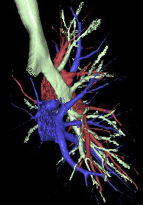
Three-dimensional computed tomography scan showing a tracheal bronchus with pulmonary vein anomalies.

Preoperative images. A, Thin-slice computed tomography (CT) showing a partly solid ground-glass opacity in the right upper lobe. (B) The apical segmental bronchus of the right lung (B1) and the posterior-anterior segmental bronchus (B2+3) are branched separately from the right main bronchus. (C) The superior branch of the pulmonary vein (V1+3) is running behind the right main pulmonary artery. (D) An aberrant pulmonary vein (V2) is running dorsally, emptying into the left atrium.

Intraoperative thoracoscopic views. The left side of the figure is the cranial side of the patient. (A) The main trunk of the pulmonary artery (PA) is taped. The white arrow shows the superior branch of the pulmonary vein (V1+3) running behind the PA. (B) The asterisk shows the aberrant V2 running in the superior segment of the lower lobe. The B1 and B2+3 have already divided. RUL, Right upper lobe; RLL, right lower lobe.

In this video, the left side of the monitor is the patient's cranial side. After opening the anterior pleura, at first, the main trunk of the pulmonary artery (PA) was exposed. The superior branch of the pulmonary vein (V1+3) was running behind the PA. The superior branch of the PA was divided by an endostapler. The middle lobe lung was partially resected to keep a surgical margin, and the residual minor fissure was divided by an endostapler. After dividing the A3 and A2b, the V1+3 was encircled and divided by an endostapler from the cranial side of the main PA. The apical segmental bronchus of the right lung (B1) was exposed and divided. Then the posterior-anterior segmental bronchus (B2+3) was divided in the same manner. The aberrant V2 was divided in conjunction with the posterior fissure by the endostapler. Video available at: https://www.jtcvs.org/article/S2666-2507(20)30395-3/fulltext .
References
-
- Barat M., Konrad H.R. Tracheal bronchus. Am J Otolaryngol. 1987;8:118–122. - PubMed
-
- Yurugi Y., Nakamura H., Taniguchi Y., Miwa K., Fujioka S., Haruki T., et al. Case of thoracoscopic right upper lobectomy for lung cancer with tracheal bronchus and a pulmonary vein variation. Asian J Endosc Surg. 2012;5:93–95. - PubMed
-
- Sumitomo R., Fukui T., Otake Y., Huang C.L. Video-assisted thoracoscopic lobectomy with an anomalous pulmonary vein. J Thorac Cardiovasc Surg. 2016;152:1398–1399. - PubMed
Publication types
LinkOut - more resources
Full Text Sources
