

doi: 10.1016/j.xjtc.2020.08.081.
eCollection 2020 Dec.
Right-to-left inverted single lung transplantation
Affiliations
- PMID: 34318084
- PMCID: PMC8307873
- DOI: 10.1016/j.xjtc.2020.08.081
Item in Clipboard
Right-to-left inverted single lung transplantation
JTCVS Tech.
.
No abstract available
Figures

Right-to-left inverted single-lung transplantation procedure via an open-space technique.

Right-to-left inverted single-lung transplantation. Video available at: https://www.jtcvs.org/article/S2666-2507(20)30557-5/fulltext .

A, Preoperative chest computed tomography scan results showed uneven disease laterality. B, Recipient pulmonary artery preparation. The pulmonary artery was divided at the periphery of A6 to leave it beyond the bronchus stump, with ligation of A3, A1+2ab, and A1+2c. The recipient pulmonary artery was cut in an oblique plane, from A1+2ab to A6, so as to obtain a wide site for anastomosis of the right-side main pulmonary artery of the donor. C, Anastomosis of the pulmonary artery was performed after making space by retraction of both the donor and recipient bronchi. D, Final image of hilar anastomosis.

Postoperative chest computed tomography scan performed 2 months after lung transplantation. Video available at: https://www.jtcvs.org/article/S2666-2507(20)30557-5/fulltext .
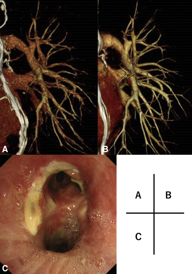
A, 3D-CT angiography image obtained at 1 week after the operation. The anastomosis site of the pulmonary artery was behind the bronchus. B, 3D-CT angiography performed 2 months later showed good patency and dilatation of the pulmonary artery and vein. C, Bronchoscopy image of anastomosis site of bronchus.
References
-
- Weder W., Inci I., Korom S., Kestenholz P.B., Hillinger S., Eich C., et al. Airway complications after lung transplantation: risk factors, prevention and outcome. Eur J Cardiothorac Surg. 2009;35:293–298. - PubMed
-
- Couetil J.-P., Argyriadis P.G., Tolan M.J., Achkar A., Carpentier A.F. Contralateral lung transplantation: a left lung implanted in the right thorax. Ann Thorac Surg. 2001;72:933–935. - PubMed
-
- van Berkel V., Guthrie T.J., Puri V., Krupnick A.S., Kreisel D., Patterson A., et al. Impact of anastomotic techniques on airway complications after lung transplant. Ann Thorac Surg. 2011;92:316–321. - PubMed
LinkOut - more resources
Full Text Sources
