Editorial
. 2021 Jan 28:7:72-78.
doi: 10.1016/j.xjtc.2020.12.042. eCollection 2021 Jun.

Reimplantation valve-sparing aortic root replacement is the most durable approach to facilitate aortic valve repair

Affiliations
Editorial

Reimplantation valve-sparing aortic root replacement is the most durable approach to facilitate aortic valve repair

Tirone David. JTCVS Tech. .

Abstract

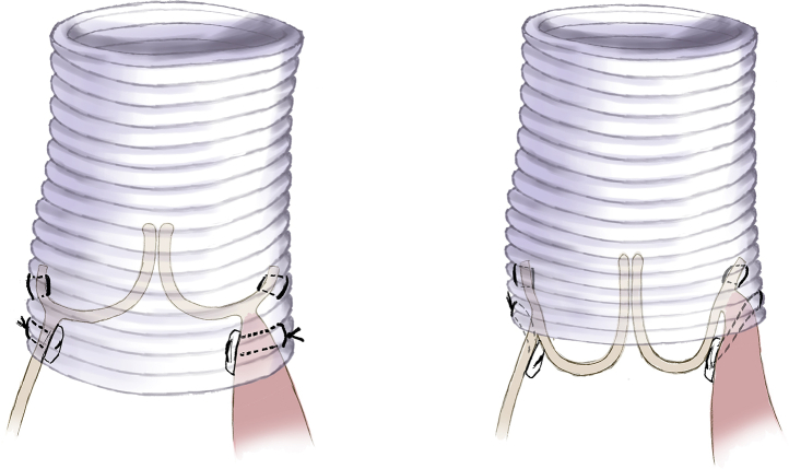
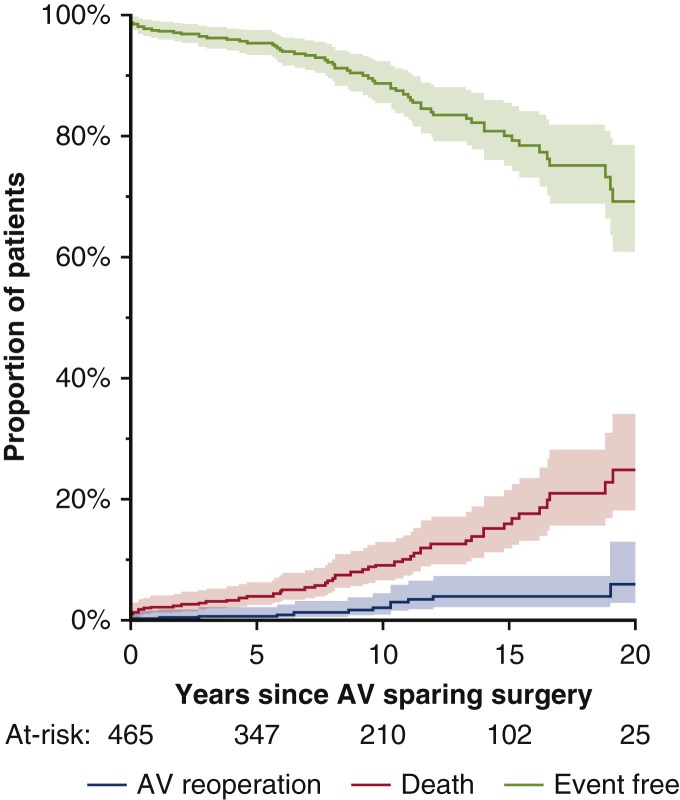
Reimplantation of the aortic valve has been performed for 3 decades, and experience shows that patient selection and meticulous operative technique are crucial to provide excellent clinical outcomes and stable aortic valve function for decades. More than the type of the Dacron graft used for the reimplantation (straight graft or Valsalva graft), we believe that attention to certain technical aspects of this operation is key to a successful and durable aortic valve reconstruction. This article describes the operative procedure as we believe it should be performed based on a learned experience with several hundred cases and summarizes the latest outcomes in a large cohort of patients followed prospectively during the past 3 decades.

Keywords: David operation; aortic insufficiency; aortic root aneurysm; aortic valve; aortic valve repair.


Figures

None
Tirone David, MD
Figure 1
Figure 1
The aortic sinuses are excised, leaving 3 to 5 mm of arterial wall attached to the aortic annulus and around the coronary artery orifices.
Figure 2
Figure 2
If a coronary artery originates close to the aortic annulus, it is safer to leave it attached to the aortic root and dissect the artery away from the aortic root and surrounding tissues (left panel). The graft is incised vertically in the area of the retained coronary artery before being secured to the left ventricular outflow tract (right panel).
Figure 3
Figure 3
The aortic annuloplasty stitches are passed from the inside to the outside of the left ventricular outflow tract through a single horizontal plane except for the area of right coronary cusp, where it may have to be placed into its subcommissural triangles (left panel). The bundle of His should be avoided and that space left without a suture. These suture line must be spatially placed in the Dacron graft using the same spatial distribution as in the left ventricular outflow tract (right panel).
Figure 4
Figure 4
Once the annuloplasty is completed, the aortoventricular junction must lay inside the Dacron graft, as illustrated in the sketch on the left panel. If the graft lies at the same level as the aortic annulus or above it as shown on the right panel, early failure is likely to occur. This is largely due to inadequate dissection of the outflow tract and placement of the sutures.
Figure 5
Figure 5
The remnants of the aortic sinuses are secured to the Dacron graft starting at the commissures and moving down toward the nadir of the annulus (left panel). This suture line must be at least 8 mm above the previous one in the left ventricular outflow tract. If a coronary artery was left attached to the commissure, the arterial wall around its orifice is sutured to the graft, as illustrated in the right panel. The incision in the graft beneath this artery should be closed with a couple of interrupted sutures.
Figure 6
Figure 6
The 3 cusps must coapt centrally inside the graft and at least 8 to 10 mm above the aortic annulus. Elongated free margins are shortened by plicating centrally along the nodule of Arantius, as shown in the upper panel. Free margin of cusps with large stress fenestration is reinforced with a double layer of 7-0 expanded polytetrafluoroethylene suture, one above and the second a millimeter or 2 beneath the free margin (lower panel).
Figure 7
Figure 7
If one or more cusps do not easily reach the central portion of the graft to coapt with the others, the intercommissural distance can be shortened by plicating the graft from the outside, as shown in the upper panel. This creates a neoaortic sinus. The complete repair is illustrated in the lower panel.
Figure 8
Figure 8
At the completion of the reimplantation procedure, transesophageal echocardiography should show the cusps entirely inside the reconstructed aortic root and the coaptation height (larger arrow) should be at greater than 8 mm and the coaptation length (smaller arrow) greater than 4 mm.
Figure 9
Figure 9
Estimates of event-free survival, death of any cause, and reoperation on the aortic valve over the years after reimplantation of the aortic valve. AV, Aortic valve.

Comment in

  • New horizons in aortic valve repair.
    Woo YJ. Woo YJ. JTCVS Tech. 2021 Apr 26;7:71. doi: 10.1016/j.xjtc.2021.04.018. eCollection 2021 Jun. JTCVS Tech. 2021. PMID: 34471910 Free PMC article. No abstract available.

References

    1. David T.E., Feindel C.M. An aortic valve-sparing operation for patients with aortic incompetence and aneurysm of the ascending aorta. J Thorac Cardiovasc Surg. 1992;103:617–622. - PubMed
    1. David T.E. Remodeling of the aortic root and preservation of the native aortic valve. Op Tech Cardiac Thorac Surg. 1996;1:44–56.
    1. David T.E. Aortic valve sparing in different aortic valve and aortic root conditions. J Am Coll Cardiol. 2016;68:654–664. - PubMed
    1. Schneider U., Aicher D., Miura H., Schäfers H.J. Suture annuloplasty in aortic valve repair. Ann Thorac Surg. 2016;101:783–785. - PubMed
    1. Lansac E., Di Centa I., Sleilaty G., Lejeune S., Berrebi A., Zacek P., et al. Remodeling root repair with an external aortic ring annuloplasty. J Thorac Cardiovasc Surg. 2017;153:1033–1042. - PubMed

Publication types

LinkOut - more resources