

Editorial
doi: 10.1016/j.xjtc.2021.01.019.
eCollection 2021 Jun.
Utility of a bespoke 3-dimensional printed model in complex transposition
Affiliations
- PMID: 34318245
- PMCID: PMC8311502
- DOI: 10.1016/j.xjtc.2021.01.019
Item in Clipboard
Editorial
Utility of a bespoke 3-dimensional printed model in complex transposition
JTCVS Tech.
.
No abstract available
Figures

Three-dimensional printed model showing a complex transposition of the great arteries.

Diagram describing the intracardiac repair. The green triangle demonstrated the ventricular septal defect (VSD) enlargement with infundibular resection and the dotted yellow line indicates the intracardiac baffle. Ao, Aorta.
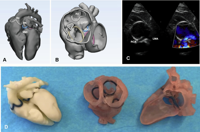
Multimodal images showing transposition of the great arteries (TGA), ventricular septal defect (VSD), small pulmonary artery (PA) and single coronary artery. A and B, reconstruction in 3 dimensions of the magnetic resonance imaging scan. C, Echocardiogram short axis showing a single coronary artery with intramural and intra-arterial trajectory. D, The 3-dimensional models. Ao, Aorta; LVOT, left ventricular outflow tract; RCA, right coronary artery; LMA, left mainstem coronary artery.

A, Postoperative echocardiographic 2-dimensional apical view, showing the intracardiac baffle, unobstructed left ventricular outflow tract, and anastomosis between pulmonary artery and the posterior wall of the aorta (Damus-Rastelli). B, Magnetic resonance imaging single-shot steady-state free precession images in sagittal view showing the right ventricle-pulmonary artery conduit (left panel), the Damus-Rastelli (central panel), and the ascending aorta (right panel). Ao, Aorta; PA, pulmonary artery; LVOT, left ventricular outflow tract.

Three-dimensional reconstructed image showing complex transposition of the great arteries. A, External view, B, With anterior wall of the right ventricle removed. Ao, Aorta; PA, pulmonary artery; VSD, ventricular septal defect.
References
-
- Honjo O., Kotani Y., Bharucha T., Mertens L., Caldarone C.A., Redington A.N., et al. Anatomical factors determining surgical decision-making in patients with transposition of the great arteries with left ventricular outflow tract obstruction. Eur J Cardiothorac Surg. 2013;44:1085–1094. - PubMed
-
- Kalfa D., Vergnat M., Baruteau A.E., Belli E. Damus anastomosis associated with REV/Rastelli procedure allows to extend indications for anatomical repair in complex transposition of great arteries. Interact Cardiovasc Thorac Surg. 2014;18:844–846. - PubMed
-
- Ceithaml E.L., Puga F.J., Danielson G.K., McGoon D.C., Ritter D.G. Results of the Damus-Stansel-Kaye procedure for transposition of the great arteries and for double-outlet right ventricle with subpulmonary ventricular septal defect. Ann Thorac Surg. 1984;38:433–437. - PubMed
-
- Kumar T.K.S., Amin N., Sathanandam S., Knott-Craig C.J. Management of coronary artery arising from nonfacing sinus in transposition of great arteries. J Thorac Cardiovasc Surg. 2018;156:e189–e190. - PubMed
Publication types
LinkOut - more resources
Full Text Sources
