

Case Reports
doi: 10.1016/j.xjtc.2021.01.027.
eCollection 2021 Jun.
Spontaneous bilobar torsion managed with pneumopexy
Affiliations
- PMID: 34318276
- PMCID: PMC8311547
- DOI: 10.1016/j.xjtc.2021.01.027
Item in Clipboard
Case Reports
Spontaneous bilobar torsion managed with pneumopexy
JTCVS Tech.
.
No abstract available
Figures
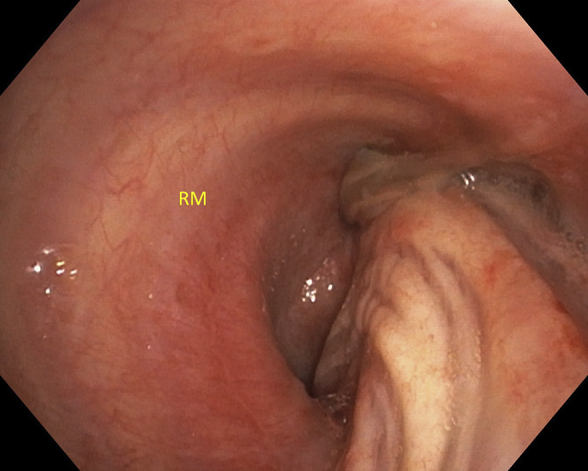
Distorted right mainstem (RM) bronchus without visualization of the right upper lobe bronchus.
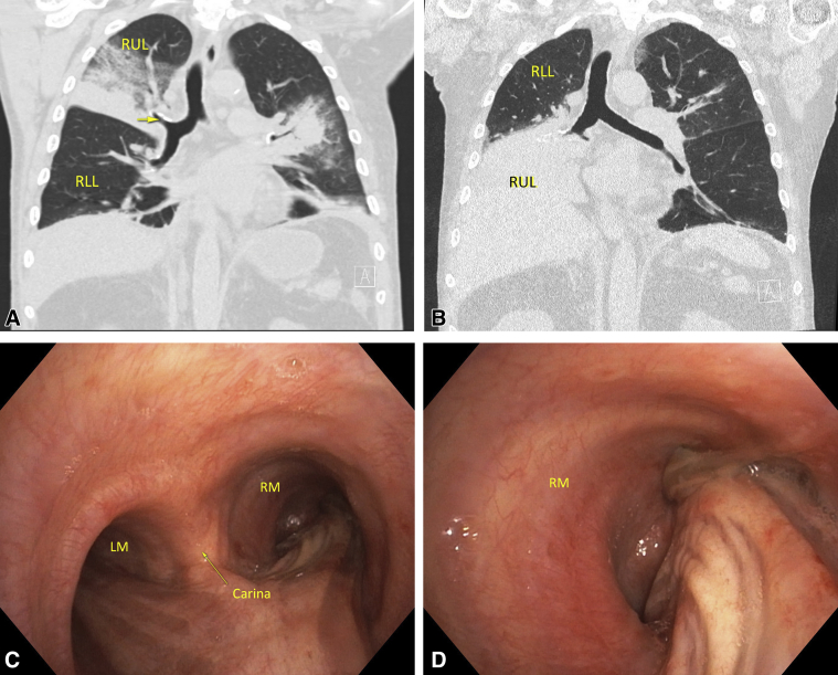
A, Initial admission computed tomography (CT) scan showing a coronal view of the right lung with right upper lobe (RUL) consolidation. The arrow is pointing to the RUL bronchus. B, CT 2 days after admission showing the right lower lobe (RLL) at the apex of the right chest and the consolidated RUL inferiorly and possibly right middle lobe (RML). Note the displacement of the RUL bronchus. C, Fiberoptic bronchoscopy demonstrating distal trachea, carina, left mainstem (LM) bronchus, and right mainstem (RM) bronchus. D, Distorted RM bronchus without visualization of the RUL bronchus.
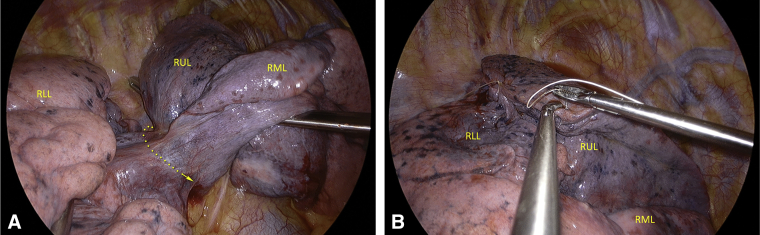
A, Thoracoscopic view demonstrating atelectatic right upper lobe (RUL) and right middle love (RML) with a narrow common bronchovascular pedicle. B, Suture pneumopexy of RUL and right lower lobe (RLL).
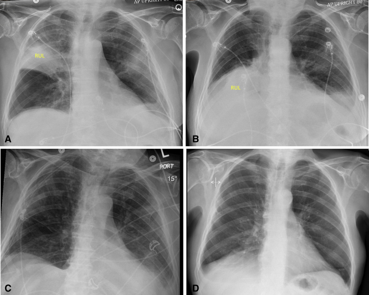
Chronology of chest radiographs from admission to discharge. A, Admission chest radiograph from the outside hospital demonstrating right upper lobe (RUL) consolidation. B, Interval chest radiograph showing migration of the RUL consolidation to right lower lung field. C, Immediate postoperative chest radiograph after thoracoscopic exploration. D, Postoperative chest radiograph 3 weeks after discharge from the hospital.
References
-
- Cable D.G., Deschamps C., Allen M.S., Miller D.L., Nichols F.C., Trastek V.F. Lobar torsion after pulmonary resection: presentation and outcome. J Thorac Cardiovasc Surg. 2001;122:1091–1093. - PubMed
-
- Hennink S., Wouters M.W.J.M., Klomp H.M., Baas P. Necrotizing pneumonitis caused by postoperative pulmonary torsion. Interact Cardiovasc Thorac Surg. 2008;7:144–145. - PubMed
-
- Ohde Y., Nakagawa K., Okumura T., Kondo H. Spontaneous pulmonary torsion secondary to pseudo-Meigs' syndrome. Interact Cardiovasc Thorac Surg. 2005;4:59–60. - PubMed
-
- Raynaud C., Lenoir S., Caliandro R., Raffenne L., Validire P., Gossot D. Spontaneous middle lobe torsion secondary to pleural effusion. Chest. 2009;136:281–283. - PubMed
-
- Moser E.S., Proto A.V. Lung torsion: case report and literature review. Radiology. 1987;162:639–643. - PubMed
Publication types
LinkOut - more resources
Full Text Sources
