A novel strategy to develop deep learning for image super-resolution using original ultra-high-resolution computed tomography images of lung as training dataset
- PMID: 34318444
- PMCID: PMC8315896
- DOI: 10.1007/s11604-021-01184-8
A novel strategy to develop deep learning for image super-resolution using original ultra-high-resolution computed tomography images of lung as training dataset
Abstract
Purpose: To improve the image quality of inflated fixed cadaveric human lungs by utilizing ultra-high-resolution computed tomography (U-HRCT) as a training dataset for super-resolution processing using deep learning (SR-DL).
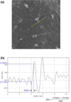
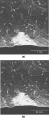
Materials and methods: Image data of nine cadaveric human lungs were acquired using U-HRCT. Three different matrix images of U-HRCT images were obtained with two acquisition modes: normal mode (512-matrix image) and super-high-resolution mode (1024- and 2048-matrix image). SR-DL used 512- and 1024-matrix images as training data for deep learning. The virtual 2048-matrix images were acquired by applying SR-DL to the 1024-matrix images. Three independent observers scored normal anatomical structures and abnormal computed tomography (CT) findings of both types of 2048-matrix images on a 3-point scale compared to 1024-matrix images. The image noise values were quantitatively calculated. Moreover, the edge rise distance (ERD) and edge rise slope (ERS) were also calculated using the CT attenuation profile to evaluate margin sharpness.
Results: The virtual 2048-matrix images significantly improved visualization of normal anatomical structures and abnormal CT findings, except for consolidation and nodules, compared with the conventional 2048-matrix images (p < 0.01). Quantitative noise values were significantly lower in the virtual 2048-matrix images than in the conventional 2048-matrix images (p < 0.001). ERD was significantly shorter in the virtual 2048-matrix images than in the conventional 2048-matrix images (p < 0.01). ERS was significantly higher in the virtual 2048-matrix images than in the conventional 2048-matrix images (p < 0.01).
Conclusion: SR-DL using original U-HRCT images as a training dataset might be a promising tool for image enhancement in terms of margin sharpness and image noise reduction. By applying trained SR-DL to U-HRCT SHR mode images, virtual ultra-high-resolution images were obtained which surpassed the image quality of unmodified SHR mode images.
Keywords: Artificial intelligence; Cadaveric lung; Convolutional neural networks; Deep learning; Ultra-high-resolution computed tomography.
© 2021. Japan Radiological Society.
Conflict of interest statement
Yoshiyuki Watanabe received a research grant from Canon Medical Systems Corp. All other authors declare that they have no conflict of interest.
Figures


References
-
- Yanagawa M, Hata A, Honda O, Kikuchi N, Miyata T, Uranishi A, et al. Subjective and objective comparisons of image quality between ultra-high-resolution CT and conventional area detector CT in phantoms and cadaveric human lungs. Eur Radiol. 2018;28:5060–5068. doi: 10.1007/s00330-018-5491-2. - DOI - PMC - PubMed
MeSH terms
LinkOut - more resources
Full Text Sources
Medical
Research Materials

