

In Vitro Glucuronidation of Caribbean Ciguatoxins in Fish: First Report of Conjugative Ciguatoxin Metabolites
- PMID: 34319092
- PMCID: PMC9215509
- DOI: 10.1021/acs.chemrestox.1c00181
In Vitro Glucuronidation of Caribbean Ciguatoxins in Fish: First Report of Conjugative Ciguatoxin Metabolites
Abstract
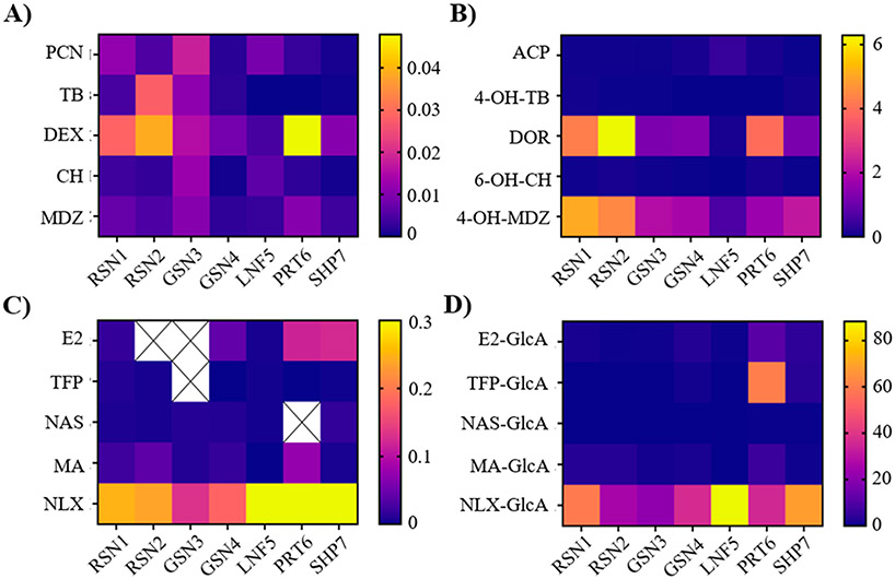
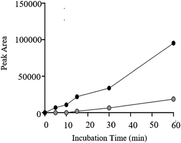
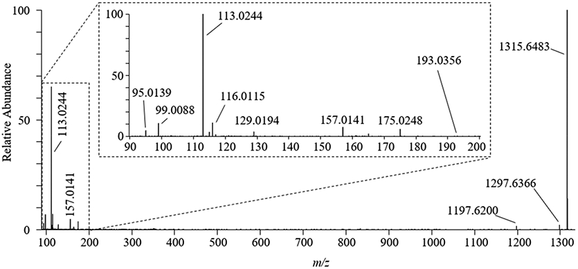
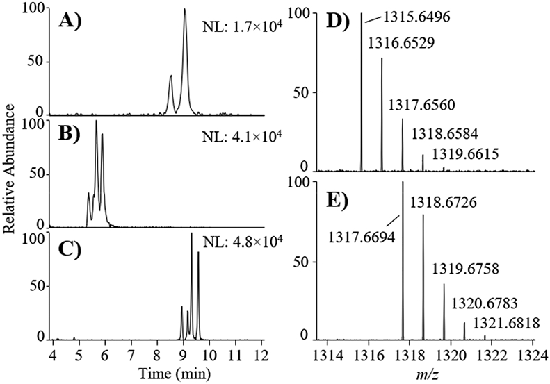
Ciguatoxins (CTX) are potent marine neurotoxins, which can bioaccumulate in seafood, causing a severe and prevalent human illness known as ciguatera poisoning (CP). Despite the worldwide impact of ciguatera, effective disease management is hindered by a lack of knowledge regarding the movement and biotransformation of CTX congeners in marine food webs, particularly in the Caribbean and Western Atlantic. In this study we investigated the hepatic biotransformation of C-CTX across several fish and mammalian species through a series of in vitro metabolism assays focused on phase I (CYP P450; functionalization) and phase II (UGT; conjugation) reactions. Using liquid chromatography high-resolution mass spectrometry to explore potential C-CTX metabolites, we observed two glucuronide products of C-CTX-1/-2 and provided additional evidence from high-resolution tandem mass spectrometry to support their identification. Chemical reduction experiments confirmed that the metabolites were comprised of four distinct glucuronide products with the sugar attached at two separate sites on C-CTX-1/-2 and excluded the C-56 hydroxyl group as the conjugation site. Glucuronidation is a novel biotransformation pathway not yet reported for CTX or other related polyether phycotoxins, yet its occurrence across all fish species tested suggests that it could be a prevalent and important detoxification mechanism in marine organisms. The absence of glucuronidation observed in this study for both rat and human microsomes suggests that alternate biotransformation pathways may be dominant in higher vertebrates.
Figures
Similar articles
-
Algal ciguatoxin identified as source of ciguatera poisoning in the Caribbean.Chemosphere. 2023 Jul;330:138659. doi: 10.1016/j.chemosphere.2023.138659. Epub 2023 Apr 10. Chemosphere. 2023. PMID: 37044143 Free PMC article.
-
Ciguatera: recent advances but the risk remains.Int J Food Microbiol. 2000 Nov 1;61(2-3):91-125. doi: 10.1016/s0168-1605(00)00382-2. Int J Food Microbiol. 2000. PMID: 11078162 Review.
-
Ciguatera Fish Poisoning in the Caribbean Sea and Atlantic Ocean: Reconciling the Multiplicity of Ciguatoxins and Analytical Chemistry Approach for Public Health Safety.Toxins (Basel). 2023 Jul 10;15(7):453. doi: 10.3390/toxins15070453. Toxins (Basel). 2023. PMID: 37505722 Free PMC article. Review.
-
Ciguatera: a public health perspective.Toxicon. 2010 Aug 15;56(2):123-36. doi: 10.1016/j.toxicon.2009.09.008. Epub 2009 Sep 23. Toxicon. 2010. PMID: 19782098 Review.
-
Liquid Chromatography Coupled to High-Resolution Mass Spectrometry for the Confirmation of Caribbean Ciguatoxin-1 as the Main Toxin Responsible for Ciguatera Poisoning Caused by Fish from European Atlantic Coasts.Toxins (Basel). 2020 Apr 21;12(4):267. doi: 10.3390/toxins12040267. Toxins (Basel). 2020. PMID: 32326183 Free PMC article.
Cited by
-
Algal ciguatoxin identified as source of ciguatera poisoning in the Caribbean.Chemosphere. 2023 Jul;330:138659. doi: 10.1016/j.chemosphere.2023.138659. Epub 2023 Apr 10. Chemosphere. 2023. PMID: 37044143 Free PMC article.
-
3-Epimers of Caribbean ciguatoxins in fish and algae.Toxicon. 2024 Jan;237:107536. doi: 10.1016/j.toxicon.2023.107536. Epub 2023 Dec 2. Toxicon. 2024. PMID: 38043714 Free PMC article.
-
Automatic MS/MS Data Mining Strategy for Rapid Screening of Polyether Toxins Derived from Gambierdiscus Species.Anal Chem. 2025 Mar 18;97(10):5643-5652. doi: 10.1021/acs.analchem.4c06440. Epub 2025 Mar 4. Anal Chem. 2025. PMID: 40035825 Free PMC article.
-
Depuration Kinetics and Growth Dilution of Caribbean Ciguatoxin in the Omnivore Lagodon rhomboides: Implications for Trophic Transfer and Ciguatera Risk.Toxins (Basel). 2021 Nov 1;13(11):774. doi: 10.3390/toxins13110774. Toxins (Basel). 2021. PMID: 34822558 Free PMC article.
-
Identification and cross-species comparison of in vitro phase I brevetoxin (BTX-2) metabolites in northern Gulf of Mexico fish and human liver microsomes by UHPLC-HRMS(/MS).Toxicon X. 2023 Jun 29;19:100168. doi: 10.1016/j.toxcx.2023.100168. eCollection 2023 Sep. Toxicon X. 2023. PMID: 37483846 Free PMC article.
References
-
- FAO and WHO Report of the Expert Meeting on Ciguatera Poisoning. Rome, 19–23 November 2018. Food Saf. Qual No. 9 2020, 98–101..
-
- Murata M, Legrand AM, Ishibashi Y, Fukui M, and Yasumoto T (1990) Structures and Configurations of Ciguatoxin from the Moray Eel Gymnothorax Javanicus and Its Likely Precursor from the Dinoflagellate Gambierdiscus toxicus. J. Am. Chem. Soc 112 (11), 4380–4386.
-
- Satake M, Murata M, and Yasumoto T (1993) The Structure of CTX3C, a Ciguatoxin Congener Isolated from Cultured Gambierdiscus Toxicus. Tetrahedron Lett. 34 (12), 1975–1978.
-
- Lewis RJ, Vernoux J-P, and Brereton IM (1998) Structure of Caribbean Ciguatoxin Isolated from Caranx Latus. J. Am. Chem. Soc 120, 5914–5920.
-
- Yogi K, Oshiro N, Inafuku Y, Hirama M, and Yasumoto T (2011) Detailed LC-MS/MS Analysis of Ciguatoxins Revealing Distinct Regional and Species Characteristics in Fish and Causative Alga from the Pacific. Anal. Chem 83, 8886–8891. - PubMed
Publication types
MeSH terms
Substances
Grants and funding
LinkOut - more resources
Full Text Sources
Molecular Biology Databases
Research Materials
Miscellaneous
